A 68 year-old male patient with a mass at right neck in lung TB regimen for 4 months but still weight loss and sudation. The painful mass existed for 1 month and getting bigger with skin redness.
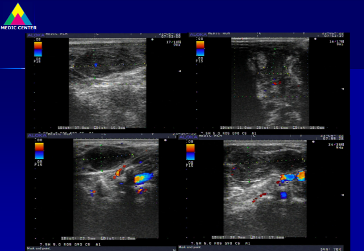
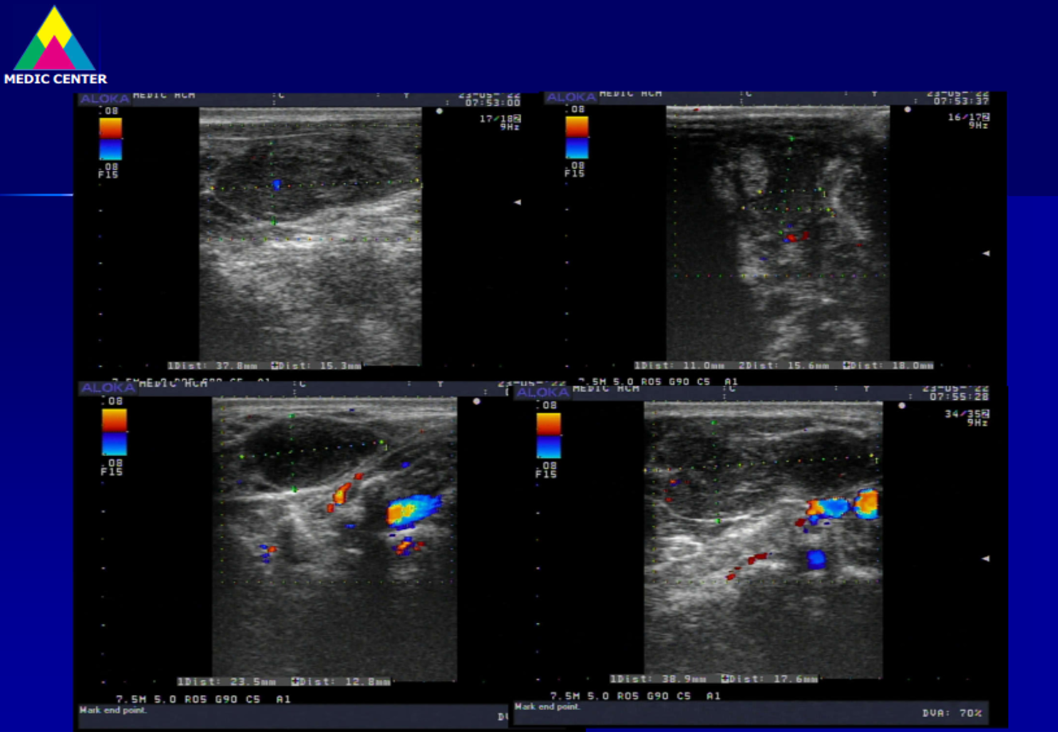
Soft tissue ultrasound detected a complexe mass #57x43 mm in muscle at right neck from angula of lower maxillary region which distorsed structure, with intramuscular fluid beside cervical vertebral column C4. It existed not any neck lymph node.


MRI confirmed a right neck tumor invasive to muscle.

Chest CT = no lung invasive, no mediastinal lymph node nor axillary node. Bone marrow biopsy exist not any malignant cell.
In surgical biopsy for chemohistopathology of the tumor resulted small cell lymphoma (C83).

The patient was treated TB lung completely and then continued lymphoma chemotherapy. Now the muscular tumor was smaller 80% and the patient remains well.
Primary muscle lymphoma is very rare entity without characteristic imaging findings but diagnostic imaging keeps a role.
REFERENCES:
Cancer Imaging (2013) 13(4), 448457 DOI: 10.1102/1470-7330.2013.0036
Imaging of musculoskeletal lymphoma
https://www.leukaemia.org.au/blood-cancer-information/types-of-bloodcancer/lymphoma/non- hodgkin-lymphoma/small-lymphocytic-lymphoma/
https://www.cancersupportcommunity.org/chronic-lymphocytic leukemiasmalllymphocytic-lymphoma
https://patientpower.info/the-curious-case-of-cll-and-sll-leukemia-lymphoma-orboth/
https://www.ncbi.nlm.nih.gov/pmc/articles/PMC6400341/
https://ashpublications.org/blood/article/131/25/2745/37141/iwCLL-guidelines-fordiagnosis- indications-for
Muscle lymphoma | Radiology Reference Article | Radiopaedia.org
Hindawi Case Reports in Radiology Volume 2017, Article ID 2068957, 7 pages
https://doi.org/10.1155/2017/2068957
Diagnostic challenge of soft tissue extranodal Hodgkin lymphoma in core-needle
biopsy: case report
No comments :
Post a Comment