Total Pageviews

Saturday 23 April 2022

CASE 631: LEFT COLON TUMOR INVADING STOMACH and CREATED a FISTULA, Dr LÂM CẨM TÚ, Dr VÕ NGUYỄN THÀNH NHÂN, Dr PHAN THANH HẢI, MEDIC MEDICAL CENTER, HCMC, VIETNAM

Female patient born in 1980,  from Bentre province went to Medic with a result of local ultrasound which described an uncertain diagnosis about an unknown mass between spleen and left kidney.

Ultrasound of Medic revealed a mass of digestive tube with pseudokidney sign and  pathologic cocard signd with its  26-33mm thick of wall that  invaded around the peritoneum on left side of abdomen. A suspection of the invaded left colon tumor was made.


MSCT confirmed the left colon tumor and revealed a connecting canal between the invaded stomach and the colon tumor.


For biopsy a gastric endoscopy was done but could not find out the gastrocolonic fistula.


Then colonic endoscopy was done in two times with results of high dysplasia of tubular adenoma.


Surgery was done to remove the left colon tumor from stomach, tail of pancreas and spleen and planned chemotherapy.


Discharge diagnosis: Adenocarcinoma of left colon grade 2  invading stomach stage 4.

CONCLUSIONS:

Young patient should have check-up whenever they can to avoid the difficult problems like that, big tumor invaded stomach, tail of pancreas and spleen that may lead to a critical operation with high risk.

Thursday 21 April 2022

CASE 630: PHYLLODES TUMOR in COVID19, Dr PHAN THANH HẢI, Dr HỒ CHÍ TRUNG, Dr JASMINE THANH XUÂN, Dr TRÂN THỊ HỒNG VÂN, MEDIC MEDICAL CENTER, HCMC, VIETNAM

 Female patient 41 yo from Baclieu province with left breast mass getting bigger for months, now after COVID 19 lockdown stop, she came to Medic to solve her big left breast.


Breast US revealed the huge left breast many structural  types solid, cystic, fatty and calcifications, but the skin and nipple were intact that was diagnosed as phyllodes tumor.


On mammography, the tumoral mass infltrated totally the left breast with calcifications. It existed benign hypersensitivity, BI-RADS 3.


MRI confirmed the huge left breast tumor.


Operation for enucleation of the left breast tumor # 1.2 kilogram from the left breast was done and, later in 3 months, will perform a cosmetic recreation of her breast.


Anapathological result was phyllodes tumor.


REFERENCE:

Cystosarcoma Phyllodes


Mary Ann Kosir

, MD, Wayne State University School of Medicine

https://www.msdmanuals.com/vi/chuy%C3%AAn-gia/ph%E1%BB%A5-khoa-v%C3%A0-s%E1%BA%A3n-khoa/nh%E1%BB%AFng-r%E1%BB%91i-lo%E1%BA%A1n-tuy%E1%BA%BFn-v%C3%BA/kh%E1%BB%91i-u-phyllodes

Monday 18 April 2022

CASE 629:Huge focal nodular hyperplasia presenting in a 7-year-old child: Dr Phan Thanh Hai, Dr Nguyen Duc Duy Linh, Dr Nguyen Ngoc Xuan Giang , MEDIC BÌNH AN, MEDIC MEDICAL CENTER, HCMC, VIETNAM


A healthy 7-year-old boy on imaging examinations, including abdominal ultrasound and computed tomography (CT):


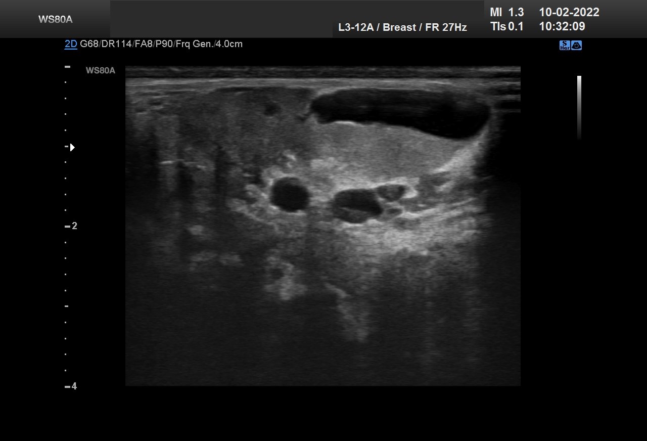
An abdominal ultrasound scan revealed a 5.3 × 5.2 cm lesion which was homogeneous  hypoechoic solid mass and its central scar in the right lobe of the liver. It existed Doppler signals of central feeding artery in the mass on a fatty infiltrated  of parenchyma of liver.


On CT  of abdomen, NECT showed a 5.7 × 5.4 cm hyperdense  mass in comparison to normal liver in the right hepatic lobe, which displayed a central star-like scar in the low-density area. On the contrast CT scan (CECT) transient, intense, and homogeneous enhancement in the hepatic arterial phase and hyperdense to normal liver in the venous phase. In the delayed phase, mass was  in hyperdense to liver while central scar was in hypodense density.


Laboratory testing revealed the following: alanine aminotransferase (ALT) and aspartate aminotransferase (AST) were 30.1 U/L (normal, 3–30 U/L) and 32.3 U/L (normal, 6–25 U/L), respectively; viral serologic tests for hepatitis B and C were negative; the alpha-fetoprotein (AFP) level was 1.6 ng/mL; and the carbohydrate antigen 19-9 (CA 19-9) level was 7.03 U/mL.


DISCUSSIONS and CONCLUSIONS

Primary tumors of the liver, including malignant and benign tumors, constitute 1%–2% of all pediatric tumors. Focal nodular hyperplasia (FNH) is a benign lesion of the liver which is usually found in healthy adults. However, FNH is rare in children, and comprises only 2% of all pediatric liver tumors. It is occasional for children suffering from FNH without symptoms. We usually use some imaging modalities (ultrasound and CT scan) to ensure the diagnosis and follow-up the FNH of liver.


References:

1. https://www.ultrasoundmedicvn.com/2017/06/case-437-liver-tumor-dr-phan-thanh-hai.html

2. https://www.ultrasoundmedicvn.com/2020/07/case-592-focal-nodular-hyperplasia-of.html