|
Man
45 yo with cough and back pain.
Chest
X-rays 1: diffuse micronodular at right/left lungs;
X-rays
2: spinal bone shows compression of
Lung
US shows thickening of pleural spaces and many B- line
signs
( US 1, US 2)
US
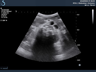
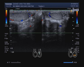
3: hypoechoic mass on the left site of paravertebral L1, and US 4: cystic mass of scrotum.
MSCT
of lung and body with CE: CT1, CT
2: micronodular lungs
CT
3 , CT 4: spine with osteolytic appearance
Radiology
report is miliary tuberculosis of the lung and Pott ‘s abscess
and scrotum abscess
Analysis
of this pus =ADA very high 63.64 ng (n<30 in pus).
Summary= It is the case of diffuse tuberculosis. |
Total Pageviews
Tuesday 29 May 2018
CASE 495: LUNG in MILIARY TUBERCULOSIS, Dr HỒ CHÍ TRUNG, Dr PHAN THANH HẢI, MEDIC MEDICAL CENTER, HCMC, VIETNAM
Tuesday 22 May 2018
CASE 494: PRIMARY LIVER LYMPHOMA (PLL),Dr PHAN THANH HẢI, MEDIC MEDICAL CENTER, HCMC, VIETNAM
Woman 62yo with 5 months of history of epigastric pain and being treated as gastritis after
gastroscopy. Ultrasound of liver reported as inhomogeneous fatty
liver.
Ultrasound
liver reviews 3 months later : US 1 manny
hypoechoic focal lesions at peripheral area
of liver with size 2-3 cm without bending vascular sign. (US 1 , US
2 CDI, US 3 central liver, US 4 liver elastography of this
hypoechoic mass is hard 41kPa, normal liver is
18kPa) US 5 : big spleen .
MSCE
with CE detected hepato slenomegaly with many nodules
captured contrast in arterial phases.
No
lymphadenomegalia in abdomen.
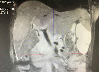
MRI
of liver with gado Images
with many hyperintense areas, T1 captured
gado enhanced peripheral ( MRI 1, 2 ,3 ,4).
Blood
tests = HBV positive EBV IGG positive Wako
test negative
Beta2
migroglobuline rised very high 8,341 UI/ IGG rised to 2,188 UI kappa IGG detected .
Summary: Based on US imaging , CT with CE, MRI with CE and blood tests
diagnosis is PLL ( primary liver lymphoma ),
wait for liver biopsy.
Sunday 13 May 2018
CASE 493: THYROID SMALL PTC, Dr PHAN THANH HẢI, MEDIC MEDICAL CENTER, HCMC, VIETNAM
Woman
52 yo, thyroid ultrasound screening detected 2 small nodules of left thyroid gland in 2015.
But now in 2018, sonologist reported back them being in TI-RADS 5, size=3.5mm. FNCA made sure that PTC.
Operation
is subtotal thyroidectomy.
See macroscopic specimen pictures.
Microscopic
report post op made sure again PTC.
Reference : medic ultrasound case 276 ptc , case 460 ptc, case 475 ptc.
Sunday 6 May 2018
CASE 492 : APPENDICULAR MUCOCELE, Dr PHAN THANH HẢI, Dr TRẦN NGÂN CHÂU, MEDIC MEDICAL CENTER, HCMC, VIETNAM.
Man 65 yo with abdomen distention (photo). For 40 years he
underwent a laparotomy in emergency by gunshot.
Ultrasound of abdomen detected at pelvis one round bordered
mass, size of 20cm. Its structure looked like cyst with many
US 1: crossed- section at middle abdomen; US 2 : with
CDI, mass no vascular inside; US 3: longitudinal scan over
MSCT scan with
CE : CT 1: this mass is cystic formation from the coecum; CT 2 : frontal view.
Appendicular mucocele
was made for diagnosing of the pelvic
mass. Operation removed one mass with mucus content from appendix.
DISCUSSION:
http://www.ytetunhantphcm.com.vn/vi/hoat-dong/khoa-hoc-dao-tao/82-ban-luan-ve-benh-u-nhay-ruot-thua-mucocele-of-the-appendix
Microscopic report is mucineous cystadenocarcinoma.
REFERENCE:
https://onlinelibrary.wiley.com/doi/pdf/10.7863/jum.2004.23.1.117
.DISCUSSION:
http://www.ytetunhantphcm.com.vn/vi/hoat-dong/khoa-hoc-dao-tao/82-ban-luan-ve-benh-u-nhay-ruot-thua-mucocele-of-the-appendix
Microscopic report is mucineous cystadenocarcinoma.
REFERENCE:
https://onlinelibrary.wiley.com/doi/pdf/10.7863/jum.2004.23.1.117
Wednesday 2 May 2018
CASE 491: TOOTHPICK MOVING TO RETROPERITONEUM, Dr PHAN THANH HẢI, MEDIC MEDICAL CENTER, HCMC, VIETNAM
Man 60 yo with epigastric pain one month ago; in emergency CT of abdomen detected a
foreign body ( FB) looked like a toothpick penetrating duodenum D2 wall.
But gastroscopy and
colonoscopy cannot find out this foreign body (FB). And so do laparoscopy later.
At Medic center, ultrasound again detected this foreign body (FB)
in retroperitoneum near IVC and aorta (US 1. US 2), very strong shadowing , US 3: longitudinal FB # 5 cm).
MSCT of abdomen non CE (CT1: crossed section this FB near
aorta , CT 2 : frontal view , CT 3: 3 D view).
Gastroscopic laparoscopy again removed this toothpick # 5 cm at the
wall of D2.
Conclusion : Toothpick can move to retroperitoneum.
REFERENCES:
REFERENCES:
In10 years at Medic it exists 5 published cases about toothpick , CASE 20 dec 2008 dr LY PHAI , MEDIC ULTRASOUND CASE 232, CASE 229, CASE 479 , CASE 491 and 7 other cases.
Subscribe to:
Posts
(
Atom
)


