Total Pageviews

Saturday 25 June 2022

CASE 640: ISOLATED COMMON ILIAC ARTERY DISSECTION, Dr Lê Văn Tài, Dr Võ Nguyễn Thành Nhân, Dr Nguyễn Tuấn Vũ, Dr Hồ Khánh Đức, Dr Phan Thanh Hải, MEDIC MEDICAL CENTER, HCMC, VIETNAM

 A  66 year-old male patient  (170 cm height, 64 kg weight) with thoracic pain was suffering from arterial hypertension [164/84 mmHg, BP 64b/min] for 3 years, and diabetes mellitus for 2 years.

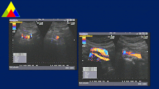
Ultrasound detected aliasing artifact from right common iliac artery [r CIA] which has been a dissection aneurysm # 34x23x20mm with 2 lumens. The peak systolic velocity (PSV) of the false lumen of right CIA was lower than the true one.  It existed atheromatic plaques in the abdominal artery [AA] wall, but it [AA] was not aneurysm. A note of arterial aneurysm dissection of the right common iliac artery [r CIA] was made.


MSCT confirmed the 19x7 mm isolated right common iliac artery dissecting.


By via percutaneous DSA repaired successfully the right common iliac artery dissection with stent.


DISCUSSION and CONCLUSIONS

Isolated common iliac artery dissection is a very rare disease which is detected by chance. The possible causes included atherosclerosis, fibromuscular dysplasia, connective tissue disease, trauma and pregnancy.

It is certainly wherever to look for an arterial aneurysm in a hypertensive old patient in routine workflow not only for abdominal aorta (AAA) but also for other arteries and its branches or inside any abdominal organes.


Friday 17 June 2022

CASE 637, 638, 639 : SECOND OPINION * X-RAYS-CT-ULTRASOUND, Dr PHAN THANH HẢI, Dr NGUYỄN VĂN CÔNG, Dr HỒ CHÍ TRUNG, Dr TRẦN NGÂN CHÂU, Dr LÊ THANH LIÊM, Dr LÝ VĂN PHÁI, Dr LÊ HỮU LINH, MEDIC MEDICAL CENTER HCMC, VIETNAM.


DISCUSSIONS

Case 1=  Pneumomediastinum

Case 2= Fracture of left clavicule

Case 3 = Sealed - off  right pleural effusion 

Ultrasound could find out the abnormalities of 3 cases above but it need to confirm the diagnosing for them by other imaging modalities.

Friday 3 June 2022

CASE 636: PARALYSIS of LEFT LARYNGEAL NERVE due to THORACIC ANEURYSM , Dr PHAN THANH HẢI, Dr ĐINH QUYẾT TÂM, MEDIC MEDICAL CENTER, HCMC, VIETNAM


A 56 yo male patient undergoes hoarse voice for 6 months after screaming, now out of breath  talking loud voice.

Laryngoscopy= Left apical arytenoid cartilage  incompletely closed.


Cardiac ultrasound  detected descending thoracic aneurysm witth aortic wall lesions.


MSCT confirmed  atresia of left vocal cord  and descending thoracic aneurysm in saccular form, non dissecting.


The recurrent laryngeal nerve RLN *from vagus nerve * supplies muscles of the larynx with the posterior and lateral cricoarytenoid.

Source **Wikipedia

In the case, descending thoracic aneurysm with aortic wall lesions may damaged the left RLN nearby which makes him the hoarse voice.


Thursday 2 June 2022

CASE 635: LEFT THORACIC WALL BULGING, Dr PHAN THANH HẢI, Dr JASMINE THANH XUÂN, Dr LÊ THANH LIÊM, MEDIC MEDICAL CENTER, HCMC, VIETNAM


  A 43 yo female patient found herself a mass of left thoracic wall for one month. It is painful when she  palpates it and moves her left arm. 

 Mammography and breast US in Vietnam detected nothing abnormal. PET-CT in Singapore was normal.


DISCUSSION and  CONCLUSION

There are some painful areas of thoracic wall that may appear in unknown microtrauma with any forces. The asymmetry of cartilage cage could be the cause of trauma in contact or due to palpation of patient herself  from her curiosity.
But in the case,  IR thermography could find out  the cause of patient complaint that noted a role of  thermography for thoracic wall bulging.