Total Pageviews

Thursday 30 December 2021

CASE 621: INCIDENTAL THYROID SMALL TUMOR, Dr PHAN THANH HẢI, Dr NGUYỄN TUẤN CƯỜNG, MEDIC MEDICAL CENTER, HCMC, VIETNAM

Female patient 48 yo came to Medic Center for a check-up .

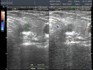
Ultrasound for thyroid incidentally detects a small nodule in left lobe that belongs TI-RADS 5 classification. Solid nodule, not well limited border,  hypoechoic, non hypervascularized without calcification. 

Lab data  TSH usensitive 1.4ulU/mL, free T4 1.13ug/dL, Antithyroglobulin 498UI/mL, Anti Microsomal [TPOAb] 37.47UI/mL.

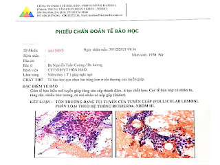
Although there is guidelines of no need to biopsy for thyroid nodule under 1 cm of diameter, a FNAC was performed. 


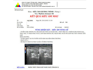
And  histopathological report is a follicular lesion, BETHESDA System Group III that means a follow-up planning must be made and in some day removing small tumor will be done.


Monday 27 December 2021

CASE 620: RECURRENT TONGUE CANCER, Dr PHAN THANH HẢI, Dr VÕ NGUYỄN THỤC QUYÊN, MEDIC MEDICAL CENTER, HCMC, VIETNAM

Female patient 48 yo suffers from a tumor of her tongue for 2 months. The tongue tumor maybe is recurrent from the small tongue tumor  3 years before and now is still a scar on right border of tongue, she said. Some lymph nodes were removed and she underwent radiation therapy on neck region.


Ultrasound detects a hypoechoic mass at right border of tongue, but is more deeper and larger than its surface. # 41x40mm. 


Later MRI confirmed the tongue tumor at right border but it is still one side and not comes to over the midline of the tongue.


Partial tongue was removed and reconstructed to keep her normal voice. Report of histopathology is squamous cell carcinoma, grade II.


Monday 20 December 2021

CASE 619 : ABDOMINAL AORTIC DILATATION, Dr PHAN THANH HẢI, Dr TRẦN THỊ THANH NGA, Dr VÕ NGUYỄN THÀNH NHÂN, Dr NGUYỄN THÀNH ĐĂNG, MEDIC MEDICAL CENTER, HCMC, VIETNAM

Old male patient 70 yo, with  AAA suspected came to Medic for reexamination.

Abdominal CT with contrast thought about subrenal non dissection AAA, diameter # 29x32mm, with intramural aortic thrombus and  aortic wall plaques. Left iliac artery in dilatation with plaque and ulcer of vascular wall.


But Doppler color ultrasound showed a dilatation of subrenal abdominal aorta  # 60x18mm with thombus that narrows 30% of  lumen and aliasing artifacts into.


Later MRI of abdomen without Gadolinium confirmed a dilatation of abdominal aorta in 2 sections, the last one with plaques in subrenal part of aorta . Left commun iliac artery with plaques is also in dilatation.


MRI of Medic Center could perform vascular imaging without Gadolinium enhancement.