Total Pageviews

Sunday 20 March 2022

CASE 628: HYDROCELE VAGINALIS, Dr PHAN THANH HẢI, Dr NGUYỄN MINH THIỀN, MEDIC MEDICAL CENTER, HCMC, VIETNAM

 Male patient was born in 1937, with diabetis and arterial hypertension for 10 years. Getting big scrotum for 30 years and had been withdrawed 3 times but he still has feeling discomfort at scrotum since 6 years.


Ultrasound revealed amount of fluid into scrotum that was more in left side.


CT scan comfirmed hydrocele vaginalis more  on left side than right one.


Under guiding ultrasound,  an amount of 350 mL yellowish fluid was withdrawed from left scrotum.


CONCLUSIONS

Choosing the fluid withdrawal technique under controlled  ultrasound for this case is reasonable as hypertension and diabetic status of the patient.

REFERENCE


Sunday 13 March 2022

CASE 627: PROSTATE CANCER with SHARING SHEAR WAVE ELASTOGRAPHY, Dr PHAN THANH HẢI, NGUYỄN MINH THIỀN, MEDIC MEDICAL CENTER, HCMC, VIETNAM

 Male patient was born in 1940,  with difficult urine discharge and high level of PSA > 100 ng/mL.

His prostatic hypertrophy with  10-30 mm lesions were in 2 lobes, in clasifying PI-RADs 5.

Seminal vesicules were invaded. Pelvic nodes 10-12 mm. Renal cysts of 2 kidneys. Right kidney  hydronephrosis degree I.  Bone metastasis vertebral column [thoracic, sacrolumbar], pelvic and femoral bones.


Enlarged prostate, structure change, not clear limited between transitional and peripheral zones. Hypervascular Doppler signals. Irregular prostatic capsule infiltrating around.


STRAIN ELASTOGRAPHY


DISCUSSIONS
CONCLUSION


Friday 11 March 2022

CASE 626: T-SHAPE IUD PENETRATING UTERUS TO URINARY BLADDER, Dr PHAN THANH HẢI, LÊ THỐNG NHẤT, JASMINE THANH XUÂN, VÕ NGUYỄN THÀNH NHÂN, MEDIC MEDICAL CENTER, HCMC, VIETNAM.

 Female patient 46 yo  with trouble of urine discharge for 1 month.

She wore her IUD for 8 years but could not find  it out 4 years before in giving up this contraception measure.

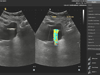
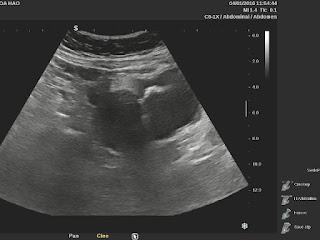
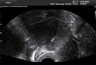
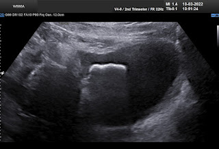
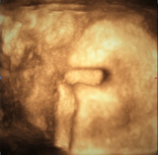
Transabdominal ultrasound and TVS revealed a metallic foreign body in her bladder with strong color comet tail artifact.


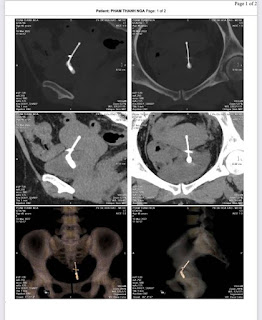
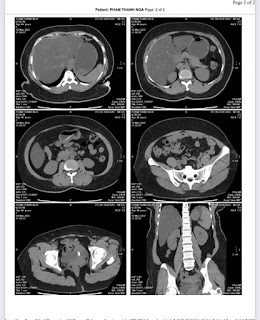
And later MSCT confirmed the T- shape IUD which penetrated to urinary bladder and was coated outside surface of it in formation of stone.


Endoscopic surgery successfully performed on March 15 to remove the stone made T- shape IUD in TD hospital.


Through anterior face of uterus, the  body and one branch of T-shape IUD migrated inside the urinary bladder that a part of it was covered by stone.  Another branch of the IUD has been in the muscular layer of the urinary bladder that  adhered the urinary bladder wall to the uterus.


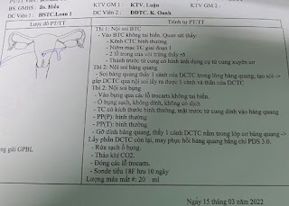
Endoscopy in surgery showed stone in urinary bladder. 


Specimen of  the T-shape IUD with stone covered on part, and another broken branch was in uterine muscle that adhered urinary bladder.

CONCLUSIONS:

Migration of T-shape IUD has highly risk of penetration the  hollow organes like rectum, urinary bladder. Ultrasound may help to detect the ectopic T-shape IUD but it needs obviously using other diagnostic imaging modalities,  endoscopic tools to confirm the status and location of it for appropriate management to the patient. 


REFERENCE

CASE 514 VUD