A 46 yo female patient goes to Medic center in breast ultrasound screenning.
Breast ultrasound detects an [4x10mm] echo mass, irregular border, inclined axis, with microcalcifications on the right breast.
The right breast mass comes from a tubular breast with microcalcifications inside.
There is not vascular signal in the right breast mass on Doppler ultrasound.
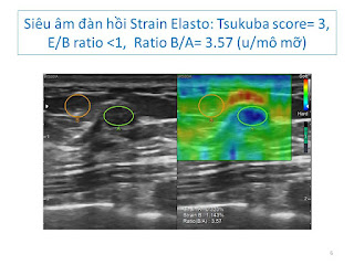
Elastoultrasound strain score 3, ratio B/A=3.57.
Mammography= On right breast it exists a mass # 10 mm, high density, blurre border with microcalcication foci ingathering.
Breast MRI with gado= Mass of right breast with high signal on T2W2 and low on T1W1, non captured CE, and some breast cysts both 2 sides.
Axillary lymph nodes are inflammed nodes.
Breast thermography: Nothing abnormal detected, due to it is a small tumor.
Result of core biopsy of the right breast tumor= Invasive breast carcinoma of no special type, grade 2.
Small size breast tumor <10mm may be revealed early in yearly screenning.
Size, location, characteristic findings will be informed with multimodalities of diagnostic imaging= ultrasound, MRI, thermography and core biopsy.
Pathohistological result is appropriate evident for breast tumor diagnosing.


No comments :
Post a Comment