Total Pageviews

Friday 11 March 2022

CASE 626: T-SHAPE IUD PENETRATING UTERUS TO URINARY BLADDER, Dr PHAN THANH HẢI, LÊ THỐNG NHẤT, JASMINE THANH XUÂN, VÕ NGUYỄN THÀNH NHÂN, MEDIC MEDICAL CENTER, HCMC, VIETNAM.

 Female patient 46 yo  with trouble of urine discharge for 1 month.

She wore her IUD for 8 years but could not find  it out 4 years before in giving up this contraception measure.

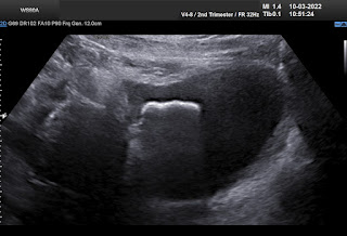
Transabdominal ultrasound and TVS revealed a metallic foreign body in her bladder with strong color comet tail artifact.


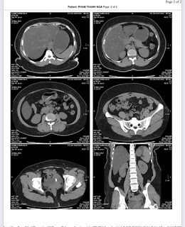
And later MSCT confirmed the T- shape IUD which penetrated to urinary bladder and was coated outside surface of it in formation of stone.


Endoscopic surgery successfully performed on March 15 to remove the stone made T- shape IUD in TD hospital.


Through anterior face of uterus, the  body and one branch of T-shape IUD migrated inside the urinary bladder that a part of it was covered by stone.  Another branch of the IUD has been in the muscular layer of the urinary bladder that  adhered the urinary bladder wall to the uterus.


Endoscopy in surgery showed stone in urinary bladder. 


Specimen of  the T-shape IUD with stone covered on part, and another broken branch was in uterine muscle that adhered urinary bladder.

CONCLUSIONS:

Migration of T-shape IUD has highly risk of penetration the  hollow organes like rectum, urinary bladder. Ultrasound may help to detect the ectopic T-shape IUD but it needs obviously using other diagnostic imaging modalities,  endoscopic tools to confirm the status and location of it for appropriate management to the patient. 


REFERENCE

CASE 514 VUD

Saturday 26 February 2022

CASE 625: HEPATIC PREGNANCY, Dr PHAN THANH HẢI, Dr PHẠM VIỆT THANH, TỪ DỤ HOSPITAL , MEDIC MEDICAL CENTER, HCMC, VIETNAM

An intrahepatic pregnancy case in over 4 months could not diagnose by transvaginal ultrasound [TVS] for a 27yo patient, PARA 1001, beta HCG elevated in Kien giang province.

2 months before, on June 13, 2007, suspected an ectopic pregnancy, the patient had been removed her right ovarian tube although not finding out the gestational sac in Kien giang.

 But, in 2 months later, with her RUQ pain from August 20, 2007, abdominal ultrasound at Medic Center and Tu Du hospital HCMC detected a 23 weeks fetus living in liver with positive heartbeats.


Placenta adhered largely to liver

On August 28, 2007, CT confirmed hepatic pregnancy aged 23 weeks that was entirely inside right lobe of liver. MSCT Angio with CE revealed vessels of fetal bag which was from hepatic artery and placenta largely adhered to liver.


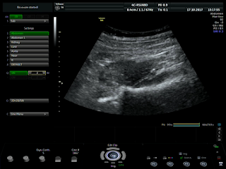
Ultrasound and abdominal CT in Medic Center detected a fetus # 23 weeks in the abdomen below the right diaphragm and inside right lobe of liver. The dimensions of fetal mass were 12x15x17cm. The placenta was 47mm thick, invaded the right liver and having a vascular supply from the right hepatic artery.

Diagnosis: great subhepatic pregnancy, indication for surgery and poor prognosis of critical case.

It would be a difficult case with high risk so Tu Du Hospital  invited a surgeon from Cho Ray Hospital to perform together a surgery for the patient.

The operation removed hepatic pregnancy but placenta suddenly detached itself from liver and profuse bleeding that could not be controlled.


Surgical report:

Laparotomy on August 30, 2007, there was about 200mL of blood flowing in the abdomen. The liver surface was smooth, but there was a lumpy mass in segments VI, VII, and VIII which was covered by anterior surface of the liver.

Dissecting the anterior and inferior border of the liver, a hepatic pregnancy mass was detected inside liver. The chorionic ridges of placenta penetrated deeply into the hepatic parenchyma, attaching to 2/3 surface of the liver. 

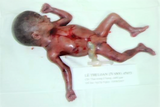
Opened the fetus bag, aspirated amniotic fluid, a dead girl fetus about 600g of weight was taken out.

Cut the umbilical cord to get the fetus and left the placenta in situ,  but the placenta detached itself from the liver, causing bleeding profusely. The surgeons had to decide to take  a part of the placenta and cut off the part of the liver that was attached to it.

This was a very complex technique and extremely difficult to stop bleeding. Tying the blood vessels from the liver stalk, the surgeons inserted gauzes to stop the bleeding. There were transfusing 8 units of blood  but it was still not controllable. About 15 minutes later, there was more bleeding from cut hepatic surface, then cardiac arrest occurred  and the patient died on surgical table.

Surgical diagnosis: Subhepatic pregnancy and death due to unstoppable bleeding.

DISCUSSIONS and  CONCLUSIONS


Hepatic pregnancy mass has grown (about 5 months # 20 - 23 weeks) in the diaphragmatic position and  had had bleeding 2 months ago, possibly from the gestational sac.

Using only the transvaginal probe,  doctors of Kien giang Province did not reveal the hepatic ectopic pregnancy and the surgeon did not find the fetus in the removed ovarian tube. Since then the fetus grew continously more 2 months and patient died due to a late ectopic pregnancy bleeding with grown fetus and placenta.

Right liver resection will be a safe solution? Currently, the technique of hemostasis of broken liver through the endovascular way is very easy to perform. 

But in this case, surgeons had controlled the main hepatic artery while  the blood loss from liver tissue and venous system continued bleeding due to the adhesion of placenta.

Recommendations: When diagnosing an ectopic pregnancy (GEU), an abdominal ultrasound examination should be using 2 probes together, vaginal probe and  abdominal probe. 

Medical treatment with MTX is safer than surgical management for early detecting ectopic pregnancy [in the abdomen, in the liver, in the spleen, ...]. 


The case was published in SIEU AM NGAY NAY 46, vol 2, 2007.


REFERENCES. 

 - PUBMED MEDLINE . Key word : hepatic ectopic pregnancy 
 - Williams Obstetrics..22nd Ed.. section III. Antepartum.. 
 - Stuart H Shippey et all Diagnosis and management of Hepatic Ectopic                    Pregnancy,   Obstet Gynecol. 2007,109 :544-6
  -Prasat K.V.S. et al.. Case report Primary hepatic pregnancy, Obgyn.net/pregnancy-birth/page=/pb/articles/hepatic-pregnancy
Wednesday 23 February 2022

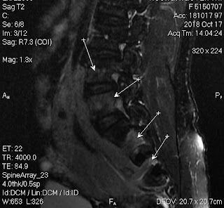
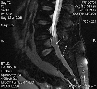
CASE 624: RIGHT PSOAS LYMPHOMA, Dr PHAN THANH HẢI, Dr LÝ VĂN PHÁI, Dr ĐẶNG VINH PHÚC, Dr VÕ NGUYỄN THÀNH NHÂN MEDIC MEDICAL CENTER, HCMC VIETNAM

Female patient 51 yo with right leg pain  and lumbago for 3 months

Lumbar spine X-Rays was normal.


Ultrasound detected  right psoas muscle poor echogeneicity like cystis pattern, no vascular, but bending aorta and right iliac artery.


Blood tests>  WBC, CRP were normal values, Beta2 Microglobuline, LDH, Ferritine were normal levels.

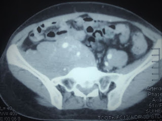
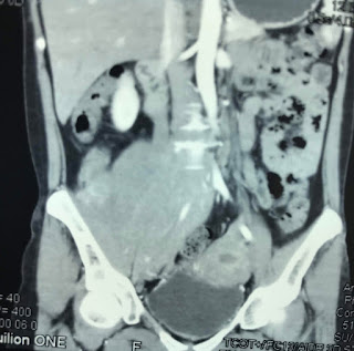
MSCT with CE= Paravertebral mass on right site, very high enhancement, deplaced iliac artery and infiltrating right psoas muscle.


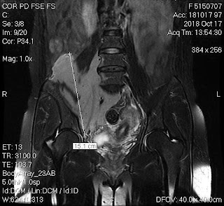
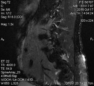
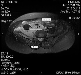
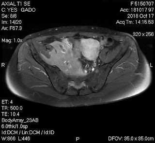
MRI with Gado= Solid mass was enrounded right psoas muscle and deplaced  right iliac artery. The tumor invaded spinal canal. Radiologist suggested retroperitoneal lymphoma.


Contrast MRI got down in urinary bladder and imaging an interesting picture of a camel like. inside urinary bladder.

Biopsy was done and result of immunohistochemistry was lymphoma B small cell.


Thursday 27 January 2022

CASE 623: PYO CHOLECYSTITIS in ELDERLY PATIENT, Dr PHAN THANH HẢI and SURGEON FELLOWS of Binh Dan Hospital, MEDIC MEDICAL CENTER, HCMC, VIETNAM.

 Male patient 83 yo with unknown cause of fever (T. 38-39 degree of Celsius) for one week. And nothing abnormal detected in clinical examination.

A full body MSCT detected a big #90x60 mm gallbladder [GB] which existed a bile debris-fluid level and #6mm thickened GB wall. Edema around GB bed was noted. A diagnosis of acute cholecystitis was done but without cause.


Later, ultrasound looks for cause of big GB status that shows bile debris of 12 mm into # 7 mm dilated GB duct and GB wall edema #16 mm. No fluid is seen around the GB. Beside GB, it exists an hepatic area of GB bed #53x28 mm which has been edema, not well limited, seems to be infiltrated and no mass effect.  Also there are some Rokitanski sinii # 35 mm in GB wall. CBD and intrahepatic bile ducts are re not dilated and without stone. An obstruction of neck of GB that causes hydrops of GB and leads to complication of acute cholecystitis with inflammation of GB bed area.


Lab data  CRP 157, WBC 10.14 [neutro 75%].

PIV antibiotics for acute cholecystitis in one week and later, laparoscopic cholecystectomy was done to remove the big gallbladder.


Macroscopic specimen of pyo cholecystitis without stone

Microscopic result is chronic cholecystitis.


Reference 

Friday 14 January 2022

CASE 622: RECTUM CARCINOMA, Dr ĐẶNG NGUYÊN KHÔI, Dr PHẠM CHÍ TOÀN, Dr VÕ NGUYỄN THÀNH NHÂN, Dr PHAN THANH HẢI, MEDIC MEDICAL CENTER, HCMC, VIETNAM

 Male patient 64yo, revealed  himself blood in stool. 

Virtual Colonoscopy MSCT detected in left pelvis a vegetative tumor of colon that narrowed 50% lumen of colon and blurred fatty tissue around.


Colonoscopy confirmed a vegetative colonic tumor which was 15 cm far from anus that made narrow lumen #50%.


Endorectal ultrasound 360 degree [EUS] detected the tumor, T3 N1 Mx. Tumor # 29x16 mm of mucosa of posterior wall invaded epithelial layer to posterior fatty tissue of rectum.


Biopsy report of the tumor is colon cancer. Differentiated carcinoma of rectum (C 18.9).


Colon rectum removed, See macro specimen.


Thursday 30 December 2021

CASE 621: INCIDENTAL THYROID SMALL TUMOR, Dr PHAN THANH HẢI, Dr NGUYỄN TUẤN CƯỜNG, MEDIC MEDICAL CENTER, HCMC, VIETNAM

Female patient 48 yo came to Medic Center for a check-up .

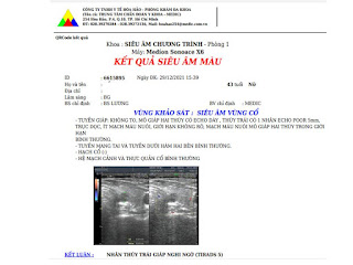
Ultrasound for thyroid incidentally detects a small nodule in left lobe that belongs TI-RADS 5 classification. Solid nodule, not well limited border,  hypoechoic, non hypervascularized without calcification. 

Lab data  TSH usensitive 1.4ulU/mL, free T4 1.13ug/dL, Antithyroglobulin 498UI/mL, Anti Microsomal [TPOAb] 37.47UI/mL.

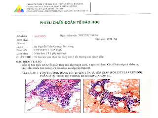
Although there is guidelines of no need to biopsy for thyroid nodule under 1 cm of diameter, a FNAC was performed. 


And  histopathological report is a follicular lesion, BETHESDA System Group III that means a follow-up planning must be made and in some day removing small tumor will be done.