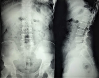
Female patient 51 yo with right leg pain and lumbago for 3 months
Lumbar spine X-Rays was normal.
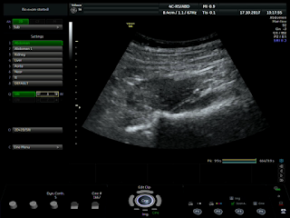
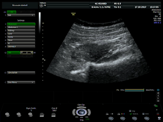
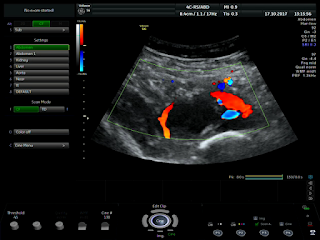
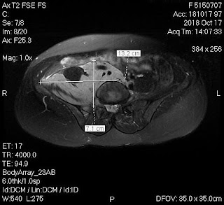
Ultrasound detected right psoas muscle poor echogeneicity like cystis pattern, no vascular, but bending aorta and right iliac artery.
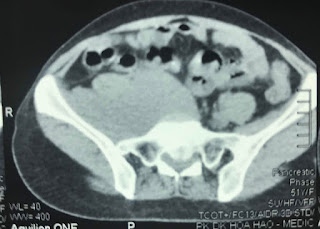
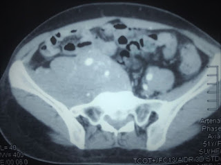
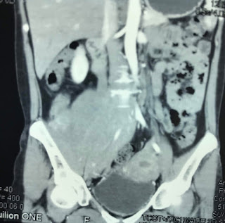
MSCT with CE= Paravertebral mass on right site, very high enhancement, deplaced iliac artery and infiltrating right psoas muscle.
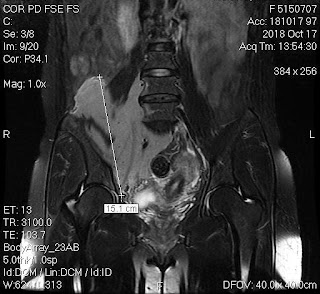
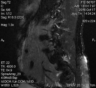
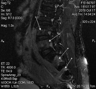
MRI with Gado= Solid mass was enrounded right psoas muscle and deplaced right iliac artery. The tumor invaded spinal canal. Radiologist suggested retroperitoneal lymphoma.
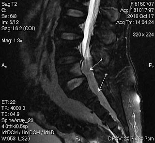
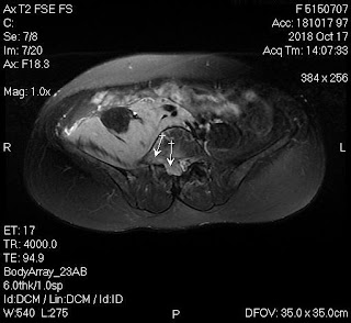
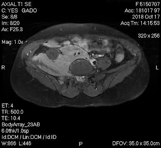
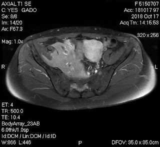
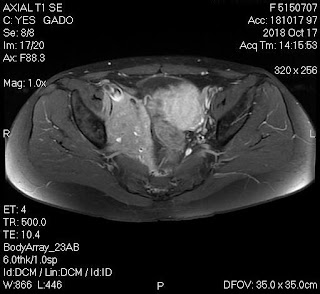
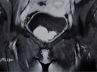
Contrast MRI got down in urinary bladder and imaging an interesting picture of a camel like. inside urinary bladder.
Biopsy was done and result of immunohistochemistry was lymphoma B small cell.

No comments :
Post a Comment