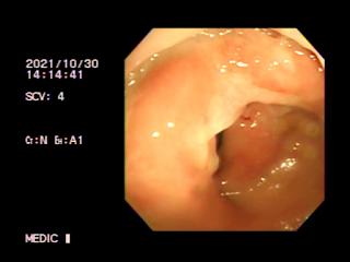
Female patient 55yo loss of weight 7 kg for 4 months with bad feelings of contracting her muscle trying to empty her bowels. There was no blood stool, but existing abnormal uterine bleeding. Digital rectal exam revealed a rigid, mobile mass at posterior wall that suggested a rectum cancer which took part of 1/3 of lumen of the rectum.
MSCT whole body detected thickening of rectum wall that adhered uterus, captured contrast and blurred fatty tissue around. Results confirmed a rectum cancer invading around with some pelvic lymph nodes.
But ultrasound and colonoscopy failed to detect the rectum tumor.
Ultrasound (TVS) only revealed uterine fibroma and cervical polyp.
DISCUSSIONS AND CONCLUSION
Clinical findings and MSCT took the clues for diagnosis of the case, but it need the concordant endoscopic result to make planning of treatment. It was difficult for endoscopy in this case, but at last it existed an evident of anapathology in the third time of endoscopy.
The female patient went through chemotherapy course and later removed rectum tumor in keeping the sphincter muscle of rectum.


No comments :
Post a Comment