Total Pageviews

Tuesday 25 September 2018

CASE 514: IUD PENETRATING TO URINARY BLADDER, Dr PHAN THANH HAI, Dr JASMINE THANH XUAN, Dr NGUYEN MINH THIEN, Dr NGUYEN TUAN VINH, MEDIC MEDICAL CENTER, HCMC, VIETNAM.


Woman 49yo with pain after urinary miction. PAST HISTORY of BEING PUT T - SHAPE IUD 20 YEARS BEFORE.
ULTRASOUND of PELVIS  DETECTED BIG URINARY BLADDER STONE ( US1)

XRAY of PELVIS  SHOWs THIS STONE  WITH IUD  INSIDE   ( X-RAYs film).

ULTRASOUND  WITH CDI  FINDs OUT TWINKLING  ARTIFACTS  WITH  COMET TAIL SIGN in GREEN AND RED COLORS ( US 2, US 3).
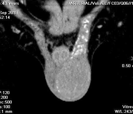
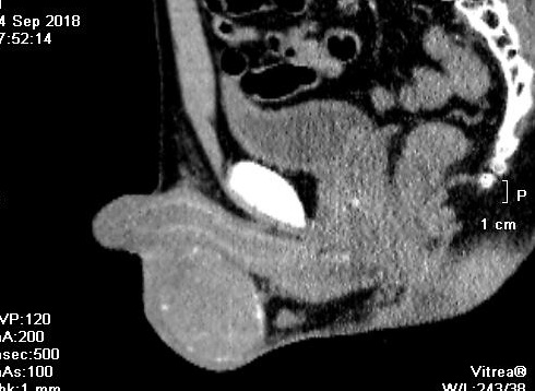


MSCT of  PELVIS  :
 CT 1=  THE  METALIC IUD  INTRA UB WALL.

CT 2:  SAGITTAL VIEW .

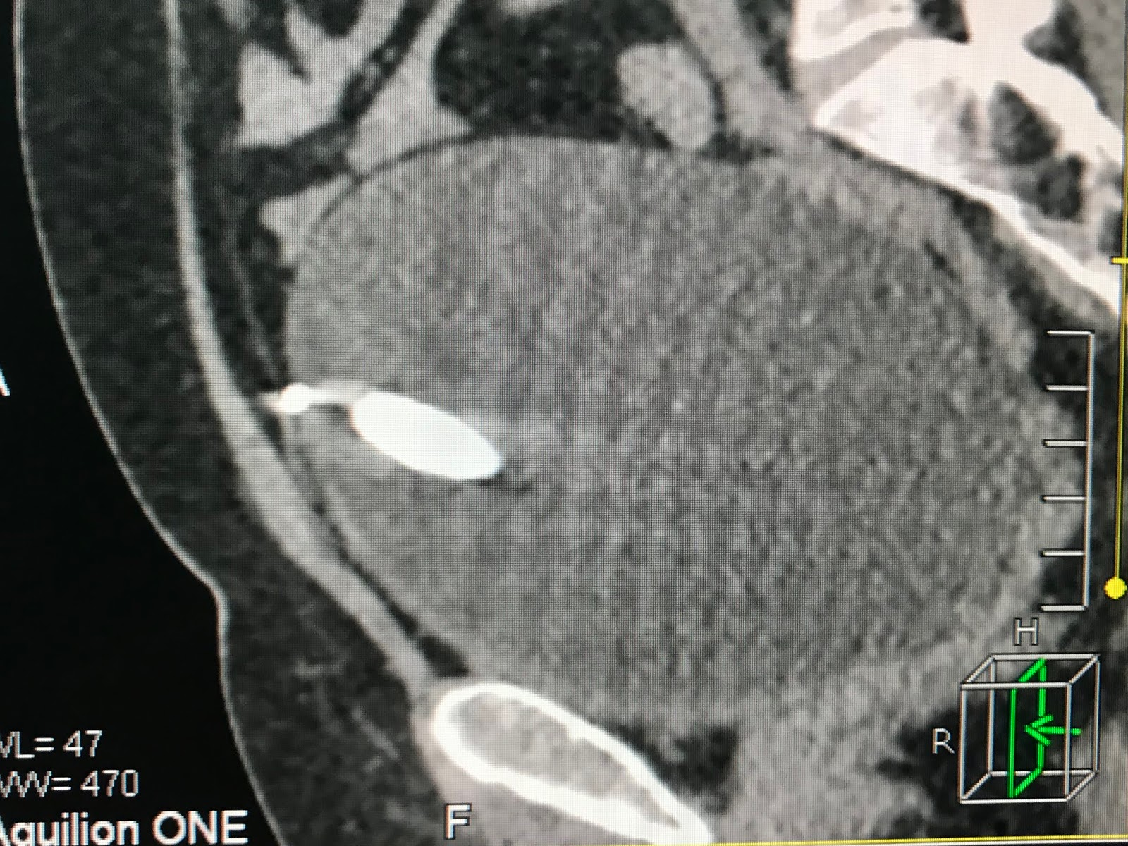
CT 3: FRONTAL VIEW : THE  IUD  INTRA UB WALL.

ENDOSCOPY DETECTED THE STONE IN VAULT OF UB.

Operation removed a big stone intra urinary bladder.


CONCLUSION =  IUD  PENETRATING TO UB WALL AND FORMATION OF STONE.

IUD Migration in MEDIC Hoa Hao

Friday 14 September 2018

CASE 513: KIDNEY TUMOR MIMICKING DROMEDARY HUMP, Dr PHAN THANH HẢI, Dr NGUYỄN MINH THIỀN, MEDIC MEDICAL CENTER, HCMC, VIETNAM.


Woman 41 yo with  righ kidney was detected abnormally  looked like dromedary  hump in general check up
Ultrasound  CDI:  US 1=  crossed section,  hypovascular pattern mass.


US 2=  longitudinal scan,  this mass liked a hump.


US 3 = elastoUS   inhomogeneous mass.


MSCT with CE, fast enhanced  contrast mass in CT 1, CT2.


MRI  with gado  shows  exophytic mass of the kidney border  (MRI 1, MRI 2).


MRI 3,4 : cystic structure  and bleeding inside.


Radiologis report is cystic tumor of righ kidney, BOSNIAK type 3 
Operation with  robot   removed  tumor in partial nephrectomy.


Operation of  this tumor  at righ kidney looked like  the seal.
 Specimen is cystic  septation,

Microspopic report is RCC.


Reference. Bosniak criteria.

Friday 7 September 2018

CASE 512: TESTICULAR TUMOR, Dr PHAN THANH HẢI, MEDIC MEDICAL CENTER, HCMC, VIETNAM.


Man 43 yo  detected left testis swollen slowly.


Ultrasound  of scrotum  finds out  left  testis too big # 4 cm x 5 cm,   solid  hypoechoic looked like a hypovascular cyst.   
US 1=  crossed section,   hypoechoic in  comparison to right testis.


US 2 = hypovascular tumor


US 3 = elastoUS,  solid tumor # 25 kPa.  


MSCT with CE   this tumor is  quick CE enhanced with calcification.
CT 1 :  frontal view


CT 2 : sagittal view


CT 3:  sagittal view of left testis tumor


Blood tests=  beta HCG , AFP = normal levels; LDH=  is 313 (n :246)
Based on clinical , ultrasound, ACD, CT  diagnostic pre op is seminoma.
Operation  removed  tumor (see macro).


Microscopic result  is  seminoma.


DISCUSSION: Why does the testis tumor being hypoechoic looked like a cyst? 

REFERENCE:


Wednesday 5 September 2018

CASE 511: LUNG CANCER,Dr PHAN THANH HẢI, MEDIC MEDICAL CENTER, HCMC, VIETNAM


Woman 60 yo with pain at left  lung after trauma. Chest x-rays detected rupture of 2 ribs
and  one mass at left lung ( see chest x-rays).


CT of  chest with CE


CT 1=   frontal view    


CT 2=  crossed section  


CT 3 = sagittal view


CT 4 with CE= very high CE enhancement.

Ultrasound at left lung  detected a tumor in invasion of the chest wall  (US1)   


US  2 = Color Doppler  shows  vascular supply to tumor   


Blood tests rised:  Cyfra 21-1= 6.02 ( N=  3.3);   CEA=  7.13  ( N= 5).

Biopsy result with CT guided is 
adenocarcinoma of the lung.


Staging of this case  with  MSCT total body detected metastasis of the brain.


Conclusion:    Lung cancer detected accidentally by trauma, stage 4.

Wednesday 29 August 2018

CASE 510: MITTELSCHMERZ SYNDROME, Dr PHAN THANH HẢI, MEDIC MEDICAL CENTER, HCMC, VIETNAM.


Woman 27 yo with  hypogastric pain. Ultrasound in emergency detected bleeding intra abdomen.
US 0=  fluid under liver area in  Morrison’s space.


US 1  = sagittal scanning at pelvis,  thickening of endometrium  and an amount of blood around pelvis.


US 2 =  transverse section at pelvis, right ovary too big  in comparison  to left one.


US 3 =  sagittal  mass at right  ovary.


Emergency blood test report  Hct 20%;  Hb 10 g/L; beta HCG  negative.


MRI 1 of abdomen detected  one mass at right  ovary.


MRI 2=  sagittal scanning of pelvis, retrouterus bleeding.


MRI 3=  frontal view of  right/left ovaries,  bleeding from right ovary.


Diagnosis is  bleeding from right ovary in Mittelschmerz syndrome.
Operation  removed 1000 ml blood clot  and right ovarian  rupture (photo) .


Mittelschmerz (German: "middle pain") is a medical term for "ovulation pain" or "midcycle pain". About 20% of women experience mittelschmerz, some every cycle, some intermittently.