Total Pageviews

Wednesday 15 August 2018

CASE 507: URACHUS TUMOR, Dr PHAN THANH HẢI, Dr LÊ VĂN TÀI, MEDIC MEDICAL CENTER, HCMC, VIETNAM.


Man 54 yo with dysuria.
Abdominal  ultrasound at hypogastric region.
US 1=  longitudinal section over  suprapubic area, reveales one mass  from  abdomen wall and connected to urinary bladder wall  at urachus site. This mass is mixed structure with cystic and solid parts.


 US 2 = crossed section of this mass.


 US 3 =Not detected any tumor in combination of 2 pictures of scanning of intraurinary bladder.


MSCT  scan of  urinary system with CE.
CT 1:  crossed-section  over urinary bladder.


CT 2:  sagittal scanning, this calcified tumor is  related to urinary bladder wall and urachus.


CT 3:  frontal view.


CT 4:  3D view of urinary system.


Radiologist report is  urachus tumor  looked like teratoma.
Operation to remove completely cystic tumor filled with mucus.


Conclusion: Ultrasound and CT make diagnostic of urachus cystic teratoma.
Pathological report is cancer of urachus tumor.


Friday 10 August 2018

CASE 506: RENAL SINUS INFLAMMATORY PSEUDO- TUMOR, Dr PHAN THANH HẢI, Prof NGUYỄN TUẤN VINH, MEDIC MEDICAL CENTER, HCMC, VIETNAM.


Man 64 yo with clinical nocturia.
Renal ultrasound detected abnormal at right kidney.
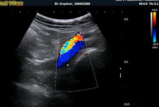
US1 : Right big renal hilus and hyperechoic.


US 2:  Crossed-section of right kidney  shows hilus vascular compression.  


US 3:   Small size of right kidney  pelvis.


US 4:   In elastographic ultrasound, right renal hilus is hard.


MRI of  abdomen.
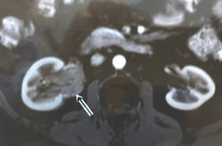
MRI 1 = Right kidney in normal size, no hydronephrosis but renal hilus is covered by one mass  look like a tumor.


MRI 2 =  Mass of  solid structure covered the right renal hilus.


MRI 3 = In frontal view, right pelvic kidney is small by this mass compressed.


Endoscopic surgery=  right pelvic kidney is  normal  without tumor.


Laparoscopy  detected  the abnormal fatty mass covered the hilus kidney. Biopsy  no tumor detected.
Microscopic report is  inflammed fatty tissue of renal hilus, that is a pseudotumor.


Reference : Hilus renal fatty stranding.


Saturday 4 August 2018

CASE 505: LEG ISCHEMIA, Dr PHAN THANH HẢI, MEDIC MEDICAL CENTER, HCMC, VIETNAM.


Man 70 yo in claudication with  pain at right leg and right foot changes in dark skin (photo).


Vascular ultrasound:   
US 1=  normal left femoral artery and vein.


 US 2=  right femoral artery and vein in stenosis.


 US 3=  in middle of right  thigh cannot find out superficial artery.


 US 4= high flow of right dorsalis pedis artery.


Thermography  shows the right leg in hypothermia.


MSCT  angio of the leg arteries:  

CT 1=  big cysts in right/left kidneys and  abdominal aorta with sclerosis plaque.


CT 2=  right  superficial femoral artery is in obstruction. 


CT 3=  small anastomosis at the level of right thigh.


Diagnosis =  obstruction of right superficial femoral artery  with many sites of deep and superficial anastomosis of femoral artery.


DSA  shows  complete obstruction of right superficial femoral artery (DSA 1)


DSA 2 = small anastomosis.


Operation removed blood clot and revascularization of the right leg. Patient remains well recovery.


Sunday 22 July 2018

CASE 504: PERIHEPATIC ABSCESS DUE TO FISHBONE, Dr PHAN THANH HẢI, Dr LÊ THỊ THANH THẢO, MEDIC MEDICAL CENTER, HCMC, VIETNAM


Woman 43 yo with  epigastric pain  has been treated as gastritis.
Ultrasound detected near  falciform ligamentun of liver  one abscess  with  foreign body long 3 cm  look like  fishbone  ( US1, US 2, US 3  SWE shows  it is very hard, US video).


CT scan no CE shows this fishbone is in a perihepatic abscess (CT1). 


One month later  the second CT examination with CE:  CT 2  shows  this abscess is  pulling abdominal wall out like a  hourglass [CT 2,  CT 3  sagital view].


Emergency operation of this abscess retrieved pus with a fishbone.


Saturday 7 July 2018

CASE 503: SUBUNGUAL GLOMUS TUMOR, Dr LÝ HỮU ĐỨC, Dr PHAN THANH HẢI, MEDIC MEDICAL CENTER, HCMC, VIETNAM.


WOMAN  46 YO  WITH  HISTORY  OF  5 YEARS AGO  PAIN AT  FIRST FINGER OF LEFT HAND, SWELLING AND BLEEDDING.
ULTRASOUND  OF THIS FINGER   WITH CDI    US 1   LONGITUDINAL SCANNING   DETECTED  ONE  MASS HYPOECHOIC BUT HYPERVASCULAR.


US 2: CROSSED-SECTION OF THIS MASS UNDER UNGUAL  
 HYPERVASCULAR  OF DISTAL FINGER.


MRI 2  =THIS MASS IS UNDER  UNGUAL   NO  INVASION OF THE BONE.


MRI 3 = CROSSED- SECTION OF THIS TUMOR IS  SUB UNGUAL WITHOUT EROSION OF THE BONE.


OPERATION  REMOVED THIS TUMOR.


 MICROSCOPIC RESULT IS  GLOMUS TUMOR.