Total Pageviews

Thursday 9 November 2017

CASE 460: CALCIFIED THYROID TUMOR , Dr PHAN THANH HẢI, Dr LÊ THANH LIÊM, Dr DƯƠNG NGỌC THÀNH, MEDIC MEDICAL CENTER, HCMC, VIETNAM


Man 52 yo, voice tone changes for  2 months, and  ENT doctor said vocal paralysis by endoscopy.
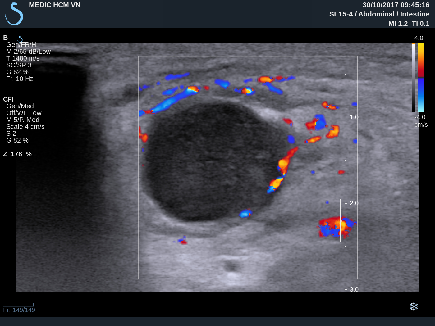
Ultrasound of the neck    
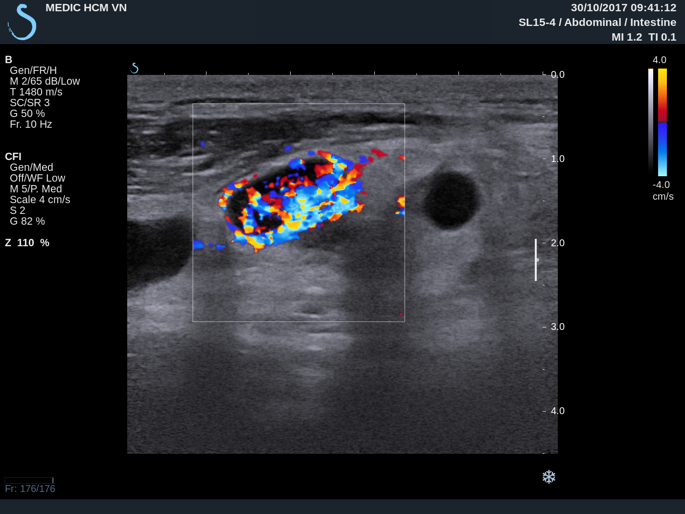
US1  left thyroid lobe normal
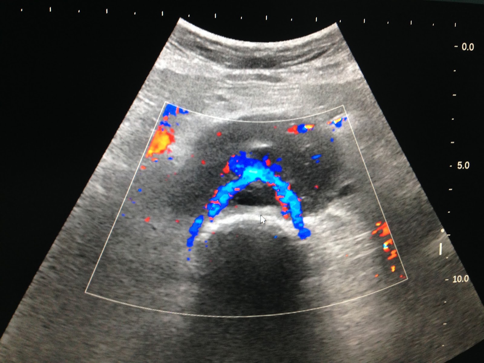
US2  right  lobe   covered by a big mass  4 cm  with strong  posterior shadowing  cannot see structure inside.


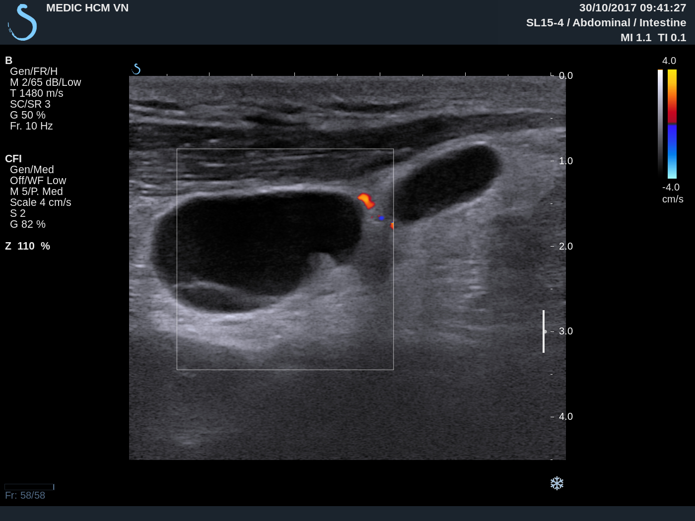
US3  near R/ CCA  small nodes with calcification #1cm.


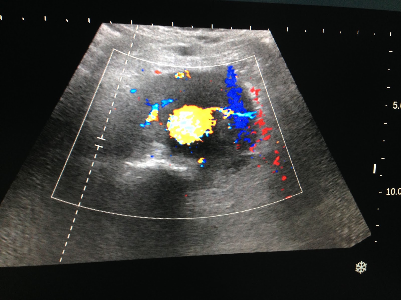
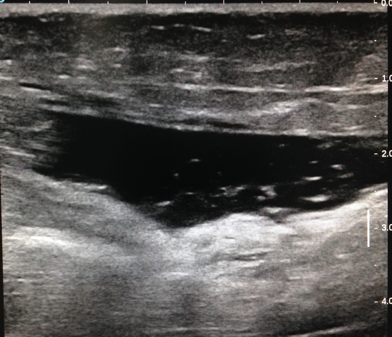
US4  with convex probe ultrasound cannot se intra tumor by very strong calcification.
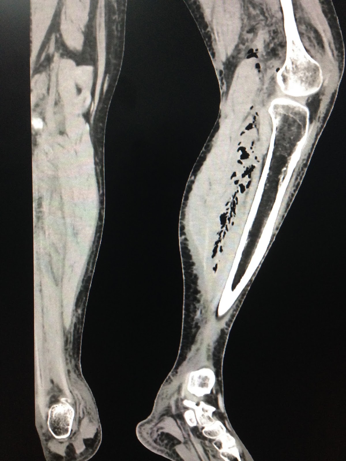
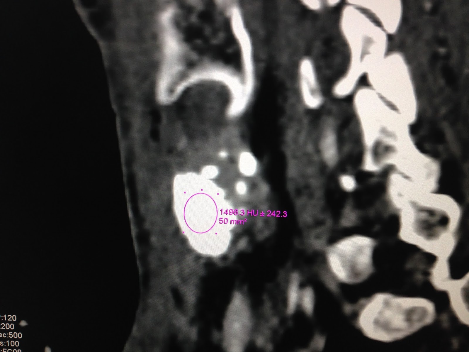

MTSC  non CE   


CT1: cross- section of the neck = mass is  very high calcification 

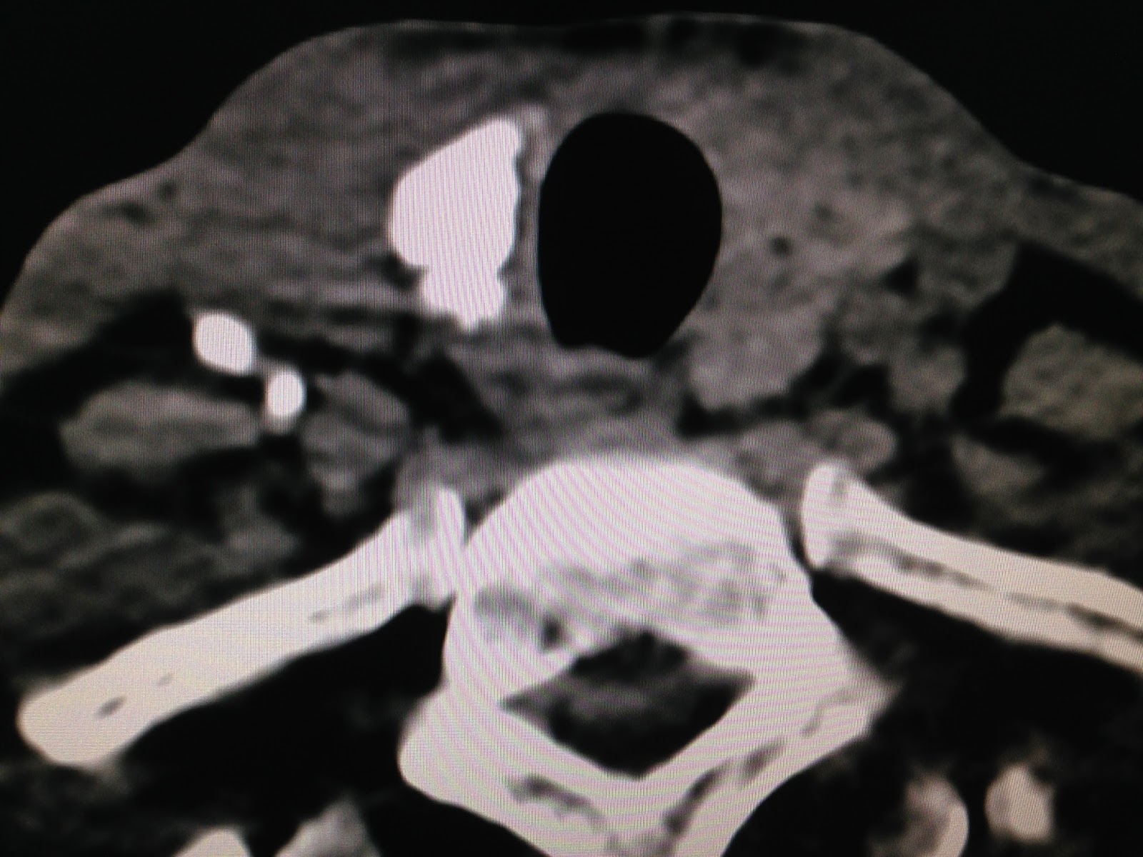
CT2 : cross- section=  calcification some lymph nodes near R/  CCA.


CT3 : frontal view  with CE=HU of this mass is  1,319 UI


CT4:   lymph node also has HU  1326UI.


CT5:  sagittal view   this mass  is  covered near the righ lobe of thyroid gland.


Blood test   TSH  is  0,041  T4  1,2   TG  97,42 (  n 3, 5-77)

 Pre-op diagnosis is thyroid cancer  metastasis neck lymph nodes.
OPERATION REMOVED RIGHT THYROID GLAND AND LYMPHADENECTOMY.

SEE  SPECEMEN 
 FOTO1    FOTO2  THYROID TUMOR CALCIFICATION


 FOTO3  LYMPH NODE


 For this case  clinical  ultrasound and CT , blood tests suggested   thyroid carcinoma   but report  of FNA  cytology is negative.

CALCITONINE = 2PG/ML   (M <18.2PG/ML)..RULES OUT MTC ( MEDULLARY THYROID CARCINOMA)
MICROSCOPIC REPORT OF SURGICAL SPECIMEN IS PAPILLARY CARCINOMA ( PTC)  METASTASIS TO SOME LYMPH NODES.


SUMMARY = PTC  WITH  HUGE CALCIFICATION  UNKNOWN.

Sunday 5 November 2017

CASE 459 : CERVICAL LYMPH NODES, Dr PHAN THANH HẢI, MEDIC MEDICAL CENTER, HCMC, VIETNAM


Man 63 yo   detected  cervical  nodules  at right neck, that were in slow growth, no pain,  no fever,   no sore throat.
Clinical palpation this  lateral nodule of the neck  from SCM chain  continuous with subclavicular group
US scan  with 12 MHz probe= thyroid gland is normal


US1: many  small 1-2 cm hypoechoic nodes ,  round border.


US2:   big node =  round,  echo very poor ,  nonvascular inside.


US3:  small node = very high vascular supply.

US4  elastoscan = very soft structure


And  the left neck is normal.

MICROSCOPIC REPORT WITH IMMUNOHISTOCHEMISTRY IS   LYMPHOMA.


Sunday 22 October 2017

CASE 458: PERIAORTIC LYMPHOMA, Dr PHAN THANH HẢI, MEDIC MEDICAL CENTER, HCMC, VIETNAM


Man 77 yo with  renal  hydronephrosis  and  insufficiency, EGFR= 11 ml/mn.
US scanning of abdomen:
US 1, US 2=2 kidneys hydronephrosis  no stone.


US 3  crossed section of aorta: Periaortic  thickening  by  hypoechoic ring.


US 4  longitudinal  scan of  abdominal aorta.
US 5: CDI.
US  6 :  scan at aorta bifurcation, CDI   longitudinal  scanning of  aorta.


CT of abdomen non CE=
CT 1=2 kidneys  hydronephrosis.
CT 2  = frontal view,   aorta is covered by the mass.
CT 3 =sagittal view.


Blood test =  betamicroglobulin 12,577UI (n=2,164)  ferritin  621ng (n =400) 

SUMMARY: 

Suspected  periaortic retroperitoneum  lymphoma in  compression of ureter  to make renal insufficiency. Wait for biopsy of  the inguinal nodes for  histo immuno stainning.

Ultrasound scanning at left inguinal region  detected many hypoechoic, hypervascular lymph nodes  that were removed for biopsy.


THE  FINAL REPORT IS FOLLICULAR LYMPHOMA WITH HISTOIMMUNO STAINING.


REFERENCE : Xin xem ca 318.


Sunday 15 October 2017

CASE 457: AML KIDNEY TUMOR, Dr PHAN THANH HẢI, Dr NGUYỄN HOÀNG ĐỨC, MEDIC MEDICAL CENTER, HCMC, VIETNAM


Man 45 yo, ultrasound screening  detected left kidney tumor.
US 1: tumor of lover pole of left  kidney,  size 4 cm,  hypoechoic pattern.
US 2:  CDI, hypovascular mass. 
US 3:  crossed section  of this left kidney tumor.
US 4:  elastoscan of this tumor is 12kPa.


MSCT with CE
CT 1:  crossed section  this tumor is low CE.
CT 2: CT density HU  is low  
CT 3:  well limited bordered tumor.


MRI  showed  the intratumoral  fatty tissue and radiologist  suggesting AML kidney tumor.


Laparoscopic operation of web resection of this tumor ( see macro1).


Microscopic result  is  AML kidney tumor.

REFERENCE:


Friday 6 October 2017

CASE 456 : LEG GAS GANGRENE, Dr PHAN THANH HẢI, MEDIC MEDICAL CENTER, HCMC, VIETNAM


Man 41 yo,with  history of liver cirhosis and ascites. 3 days fever and pain at left swollen leg.
      [Foto1]. 


    Clinical suspected lower limb DVT

  • Ultrasound  ruled out DVT ( US 1=artery and vein at left inguinal), US 2   fluid collecting 
       between muscle quadriceps; US 3  scanning at calf  detected intramuscular  air
        US 4 air and  level.


MSCT non CE detected   air in calf muscles
CT 1  crossed section;  CT 2  sagittal scanning   


Blood test, WBC  no rising;    blood culture  detected  gram  negative bacillus, 
  •  For 24 hours after  hospital admission  patient was dead with  bullous legs [Foto2].


Blood culture identifies Aeromonas caviae.
REFERENCE :