Total Pageviews

Sunday 17 December 2017

CASE 466:CHILDREN HEEL PAIN : A SEVER'S DISEASE CASE , Dr PHAN THANH HẢI PHƯỢNG, MEDIC MEDICAL CENTER, HMC, VIETNAM

9 years old male patient,  with chief complain of pain in both heels, which worsen by physical activities such as walking, running.
Physical examination: generally normal, Squeeze test (+) on right side.

 X-ray examination and ultrasound were performed.

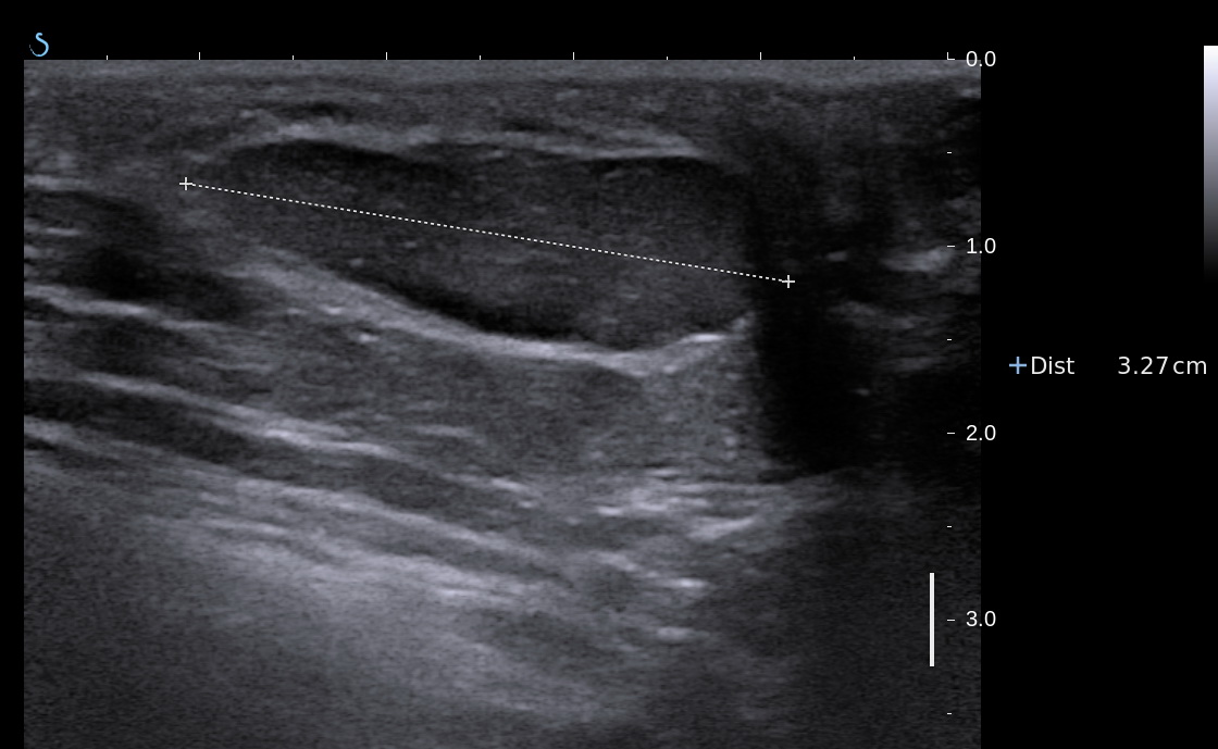
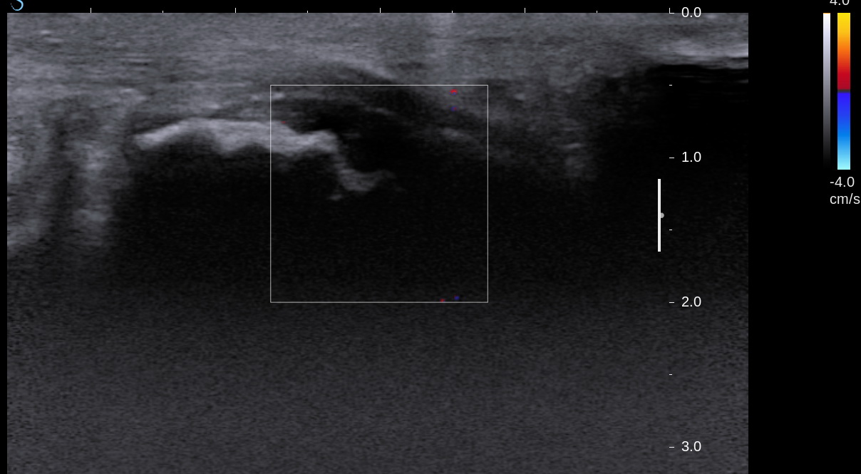
On ultrasound plantar fascia is normal. Note: the anechoic region between calcaneous is not fluid (which can indirectly suggest fascilitis in case of adult) but in fact the normal apophysis (growth plate). 


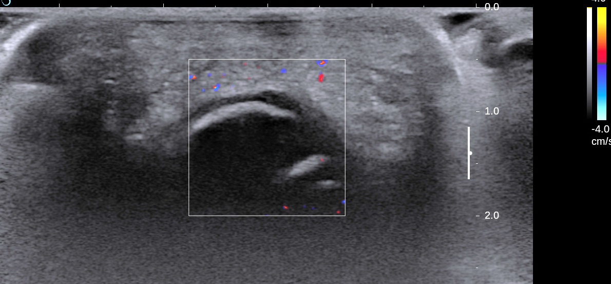
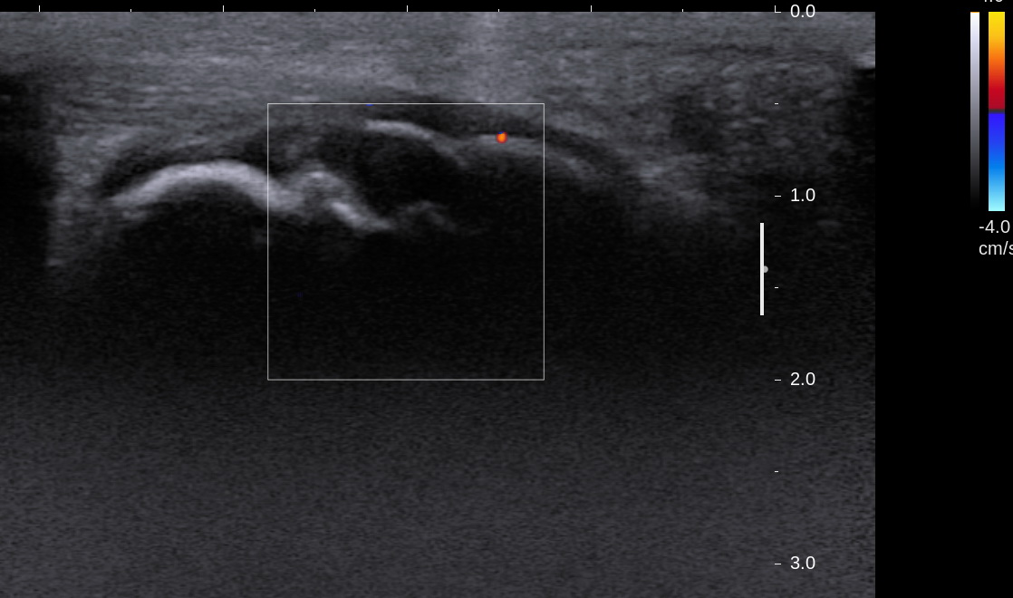
Achilles tendon is normal and remains continous fibrous echotexture (US 2), again, the rough bone surface with anechoic shown normal apophysis.

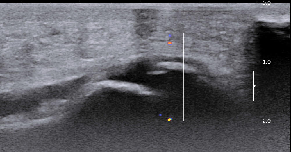
Normal distance to apophysis in both sides, no dislocation, no avulsion.


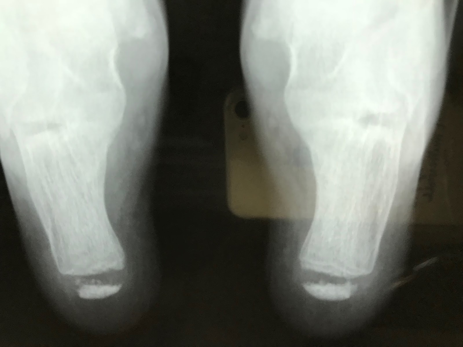
X-rays examination of both 2 heel  are normal.


Physician suggests Sever's disease, and patient was told to take some time to
rest, proper physical activity and shoes fitting.

Conclusion:
Sever's disease, the most common cause of children heel pain, known as calcaneal apophysitis is an inflammation of growth plate in heel of growing children.  Diagnosis usually bases on clinical,  and X-rays is normal. Ultrasound is suitable diagnostic tool while X-ray examination is only helpful when an ossification center of apophysis exist. Ultrasound  helps ruling out muscle strain, detect edema, lytic and avulsion.

Wednesday 6 December 2017

CASE 465: LUNG with LOFFLER SYNDROME, Dr PHAN THANH HAI. MEDIC MEDICAL CENTER, HCMC VIETNAM


Woman  45 yo with  fever and cough. Chest XRays detected many  white spots  like balloon both site the lung.


Radiologist suggested diffuse lung metastasis.
Blood tests=  WBC rise 11.27 k, eosi 20,7%,  IgE 1779 UI/mL.
Toxocara sp  positive  with od 1,809 and  all cancer marker are negative.
CT scan  with CE many opacification of  peripheral lung  booth site( CT1a/b)


CT scan the lung  with CE   the  lesion is small  regression.


Ultrasound of thorax  with small light 


After one week treatment  no fever  no cough.   Chest  Xrays  is clear.


Summary:   By the clinical, blood tests, chest XRays,  diagnosis  as  Loffler syndrome  of the lung  was made for the case.
REFERENCE


AFTER ONE MONTH  CHEST XRAY IS NORMAL  AND BLOOD TEST  STILL HIGHT EOSINOPHILE 13%  


Monday 4 December 2017

CASE 464: ANTERIOR MEDIASTINAL TUMOR, Dr PHAN THANH HẢI, MEDIC MEDICAL CENTER, HCMC, VIETNAM.


Female patient 62 yo, cardiologist send to CT scan for coronary artery  but in same time CT scanning detected one 3 cm mass  at the superior anterior madiastinal area.., well bordered.

CT1:  crossed section  non CE this mass has HU  = 54.


CT 2 :  after CE late phase HU  = 73.


CT 3 :  sagittal section, this mass is  ovoid  form at the  anterior mediastinal area.


CT 4 :  frontal view,  this mass is near aortic ascending.


CT 5 :  CE  arterial  phase,  this mass is late enhanced  and cystic formation.


Blood test is normal, and negative all cancer markers.


Radiologist suggested  thymom
Endoscopy operation of thoracotomy  removed this tumor  and  microscopic report is normal thymus tissue.
Conclusion:  it is  thymus resting.


Friday 24 November 2017

CASE 463: MADURA FOOT, Dr PHAN THANH HẢI, MEDIC MEDICAL CENTER, HCMC, VIETNAM


Man 25 yo, with  right  swollen foot   and  bleeding  after  small  trauma for  5 years.  Being treated  in many hospital  this foot  but  not better.  Biopsy 3 times in Cancer Center  with  report is chronic inflamation. (see foto 1,2).


Xray   shows  the  erosion of metatarsal bones ( xray)


Ultrasound  reported   edema with  hypervascular  soft tissue of the foot 


Biopsy of this  tumor again

Biopsy report is mycetoma.
REFERENCE:
Mycetoma

Wednesday 22 November 2017

CASE 462: LIVER ABSCESS POST ENDOSCOPIC CHOLECYSTECTOMY, Dr PHAN THANH HAI, MEDIC MEDICAL CENTER, HCMC, VIETNAM.


Man 67 y o, emergency operation by acute necrosis of  gallbladder by stone one week ago, still pain at  Murphy area. WBC = 12k with neutro 90%,  CRP= 100ng/mL.
Abdomen ultrasound detected one mass # 5 cm  at the bed of gallbladder. Mass has got fluid content  and  white structures inside with very strong  shadowing and air in formatting an abscess.
No dilatation of the biliary system.
US 1: subhepatic abscess  with strong shadowing in abscess.


US  2: umbrella sign of  the shadowing.


US 3:  elatoscan  shows  this structure is very hard.


MSCT:   CT 1:  abscess with  air and fluid filling at the bed of gall bladder which had been  removed of GB.


              CT 2:  crossed-section view of this abscess:  inhomogenous structure  and air


              CT 3:   frontal view of the abscess.

   
Radiologist reported  textilloma in suspection.


Laparoendoscopy detected an abscess in liver at the bed of gallbladder necrosis and no textilloma.

Monday 20 November 2017

CASE 461: AXILLARY MAMMARY GLAND, Dr PHAN THANH HẢI, MEDIC MEDICAL CENTER, HCMC, VIETNAM


Woman  35 yo detected  at  right axiilary  a soft  and bigger mass. Clinical looked  like a lipoma.
Ultrasound  scan of this mass :  

US 1:  longitudinal scan=  subcutaneous hypoechoic mass,  size  4cm,  well bordered.   


US 2: CDI  hypovascular  pattern.


US 3:  elastoscan of this mass = 4.3 kPa,  like fatty tissue.


MSCT  non CE:

CT 1:  frontal view of  this mass  = subcutaneous, same density of fatty tissue.


CT 2 :  zooming of this mass  showed  structure  looked like a nipple of breast.


CT 3:   crossed-sectional view of  this mass = well bodered, not connected to the right breast.


CT 4:   sagittal view of  this mass=  separation to the right breast.


Radiologist reported  an axillary  mammary gland.
Operation  for removing  this mass     (see foto  specimen),  


surgeon reported it having  fatty  and hard tissue. Microscopic report is tissue of mammary gland.


Summary of  this case:    axiilary mammary gland  mimicking as a lipoma mass.