Total Pageviews

Thursday 27 January 2022

CASE 623: PYO CHOLECYSTITIS in ELDERLY PATIENT, Dr PHAN THANH HẢI and SURGEON FELLOWS of Binh Dan Hospital, MEDIC MEDICAL CENTER, HCMC, VIETNAM.

 Male patient 83 yo with unknown cause of fever (T. 38-39 degree of Celsius) for one week. And nothing abnormal detected in clinical examination.

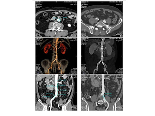
A full body MSCT detected a big #90x60 mm gallbladder [GB] which existed a bile debris-fluid level and #6mm thickened GB wall. Edema around GB bed was noted. A diagnosis of acute cholecystitis was done but without cause.


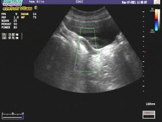
Later, ultrasound looks for cause of big GB status that shows bile debris of 12 mm into # 7 mm dilated GB duct and GB wall edema #16 mm. No fluid is seen around the GB. Beside GB, it exists an hepatic area of GB bed #53x28 mm which has been edema, not well limited, seems to be infiltrated and no mass effect.  Also there are some Rokitanski sinii # 35 mm in GB wall. CBD and intrahepatic bile ducts are re not dilated and without stone. An obstruction of neck of GB that causes hydrops of GB and leads to complication of acute cholecystitis with inflammation of GB bed area.


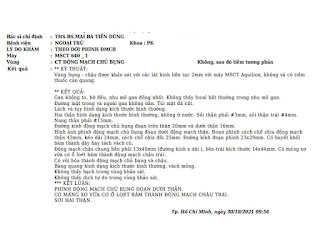
Lab data  CRP 157, WBC 10.14 [neutro 75%].

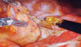
PIV antibiotics for acute cholecystitis in one week and later, laparoscopic cholecystectomy was done to remove the big gallbladder.


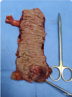
Macroscopic specimen of pyo cholecystitis without stone

Microscopic result is chronic cholecystitis.


Reference 

Friday 14 January 2022

CASE 622: RECTUM CARCINOMA, Dr ĐẶNG NGUYÊN KHÔI, Dr PHẠM CHÍ TOÀN, Dr VÕ NGUYỄN THÀNH NHÂN, Dr PHAN THANH HẢI, MEDIC MEDICAL CENTER, HCMC, VIETNAM

 Male patient 64yo, revealed  himself blood in stool. 

Virtual Colonoscopy MSCT detected in left pelvis a vegetative tumor of colon that narrowed 50% lumen of colon and blurred fatty tissue around.


Colonoscopy confirmed a vegetative colonic tumor which was 15 cm far from anus that made narrow lumen #50%.


Endorectal ultrasound 360 degree [EUS] detected the tumor, T3 N1 Mx. Tumor # 29x16 mm of mucosa of posterior wall invaded epithelial layer to posterior fatty tissue of rectum.


Biopsy report of the tumor is colon cancer. Differentiated carcinoma of rectum (C 18.9).


Colon rectum removed, See macro specimen.


Thursday 30 December 2021

CASE 621: INCIDENTAL THYROID SMALL TUMOR, Dr PHAN THANH HẢI, Dr NGUYỄN TUẤN CƯỜNG, MEDIC MEDICAL CENTER, HCMC, VIETNAM

Female patient 48 yo came to Medic Center for a check-up .

Ultrasound for thyroid incidentally detects a small nodule in left lobe that belongs TI-RADS 5 classification. Solid nodule, not well limited border,  hypoechoic, non hypervascularized without calcification. 

Lab data  TSH usensitive 1.4ulU/mL, free T4 1.13ug/dL, Antithyroglobulin 498UI/mL, Anti Microsomal [TPOAb] 37.47UI/mL.

Although there is guidelines of no need to biopsy for thyroid nodule under 1 cm of diameter, a FNAC was performed. 


And  histopathological report is a follicular lesion, BETHESDA System Group III that means a follow-up planning must be made and in some day removing small tumor will be done.


Monday 27 December 2021

CASE 620: RECURRENT TONGUE CANCER, Dr PHAN THANH HẢI, Dr VÕ NGUYỄN THỤC QUYÊN, MEDIC MEDICAL CENTER, HCMC, VIETNAM

Female patient 48 yo suffers from a tumor of her tongue for 2 months. The tongue tumor maybe is recurrent from the small tongue tumor  3 years before and now is still a scar on right border of tongue, she said. Some lymph nodes were removed and she underwent radiation therapy on neck region.


Ultrasound detects a hypoechoic mass at right border of tongue, but is more deeper and larger than its surface. # 41x40mm. 


Later MRI confirmed the tongue tumor at right border but it is still one side and not comes to over the midline of the tongue.


Partial tongue was removed and reconstructed to keep her normal voice. Report of histopathology is squamous cell carcinoma, grade II.


Monday 20 December 2021

CASE 619 : ABDOMINAL AORTIC DILATATION, Dr PHAN THANH HẢI, Dr TRẦN THỊ THANH NGA, Dr VÕ NGUYỄN THÀNH NHÂN, Dr NGUYỄN THÀNH ĐĂNG, MEDIC MEDICAL CENTER, HCMC, VIETNAM

Old male patient 70 yo, with  AAA suspected came to Medic for reexamination.

Abdominal CT with contrast thought about subrenal non dissection AAA, diameter # 29x32mm, with intramural aortic thrombus and  aortic wall plaques. Left iliac artery in dilatation with plaque and ulcer of vascular wall.


But Doppler color ultrasound showed a dilatation of subrenal abdominal aorta  # 60x18mm with thombus that narrows 30% of  lumen and aliasing artifacts into.


Later MRI of abdomen without Gadolinium confirmed a dilatation of abdominal aorta in 2 sections, the last one with plaques in subrenal part of aorta . Left commun iliac artery with plaques is also in dilatation.


MRI of Medic Center could perform vascular imaging without Gadolinium enhancement.

Friday 12 November 2021

CASE 618: SMALL UTERUS LEADS TO SUSPICION TO CEREBRAL TUMOR, Dr LE DINH VINH PHUC, Dr PHAN THANH HAI, MEDIC MEDICAL CENTER, HCMC, VIETNAM


Female tennager 13 yo from midland region of middle Vietnam go to Medic for a checkup. 

In general ultrasound  a small uterus was detected so inducing a endocrinological problem but therer is no clue about. 


For year got pain of her right leg and foot at gym without abnormal on X-ray films. From 6 months till now 3 right hand fingers, foot and fingers are involuntary in flexion and weakness feelings. Then for later 3 months her chief complaints are plenty drinking, polyuria, headache, space out, more sleeping and drowsiness. But she got no fever nor blurred vision.


MRI brain was performed and a germinoma tumor was detected above hypophysis,hypophysis and basal ganglion on right side.


Sunday 31 October 2021

CASE 617: PROSTATE on SWE TRUS with BIPLAN PROBE, Dr NGUYEN MINH THIEN, Dr PHAN THANH HAI, MEDIC MEDICAL CENTER, HCMC, VIETNAM

Male patient 64yo with fever and voiding discomfort for month.

Digital rectal examination detected a big prostate with a hard nodule on right side.

On MRI there were a 22x15mm hard node of right lobe and another one, 12x16mm, on the left of prostate which concern neoplastic foci , but  PSA=1.75 ng/mL;  F/T=20%


At Medic, an SWE elastography TRUS with biplane probe was done


On TRUS B-mode,  peripheral area of right lobe exists a 16x18 mm hypoechoic nodule, not well-limited,  hypervascular and irregular capsule of right lobe.

On strain elastography, nodule is harder than transitional area while surrounded area of right lobe is softer than the nodule.

On 2D SWE, stiffness of nodule on right side has got a value of 50 kPa [mild hard] while lesion of transitional area of left lobe of 47 kPa.


By via  transperineal a biopsy of prostate was perfomed and histopathological result is TB of prostate and subacute prostatitis.


Conclusion: 

SWE TRUS with biplan probe helps imaging lesions of prostate with more  information to plan for treatment better.