Total Pageviews

Tuesday 12 March 2019

CASE 541: RIB TUMOR, Dr PHAN THANH HAI, Dr HO CHI TRUNG, MEDIC MEDICAL CENTER, HCMC, VIETNAM.


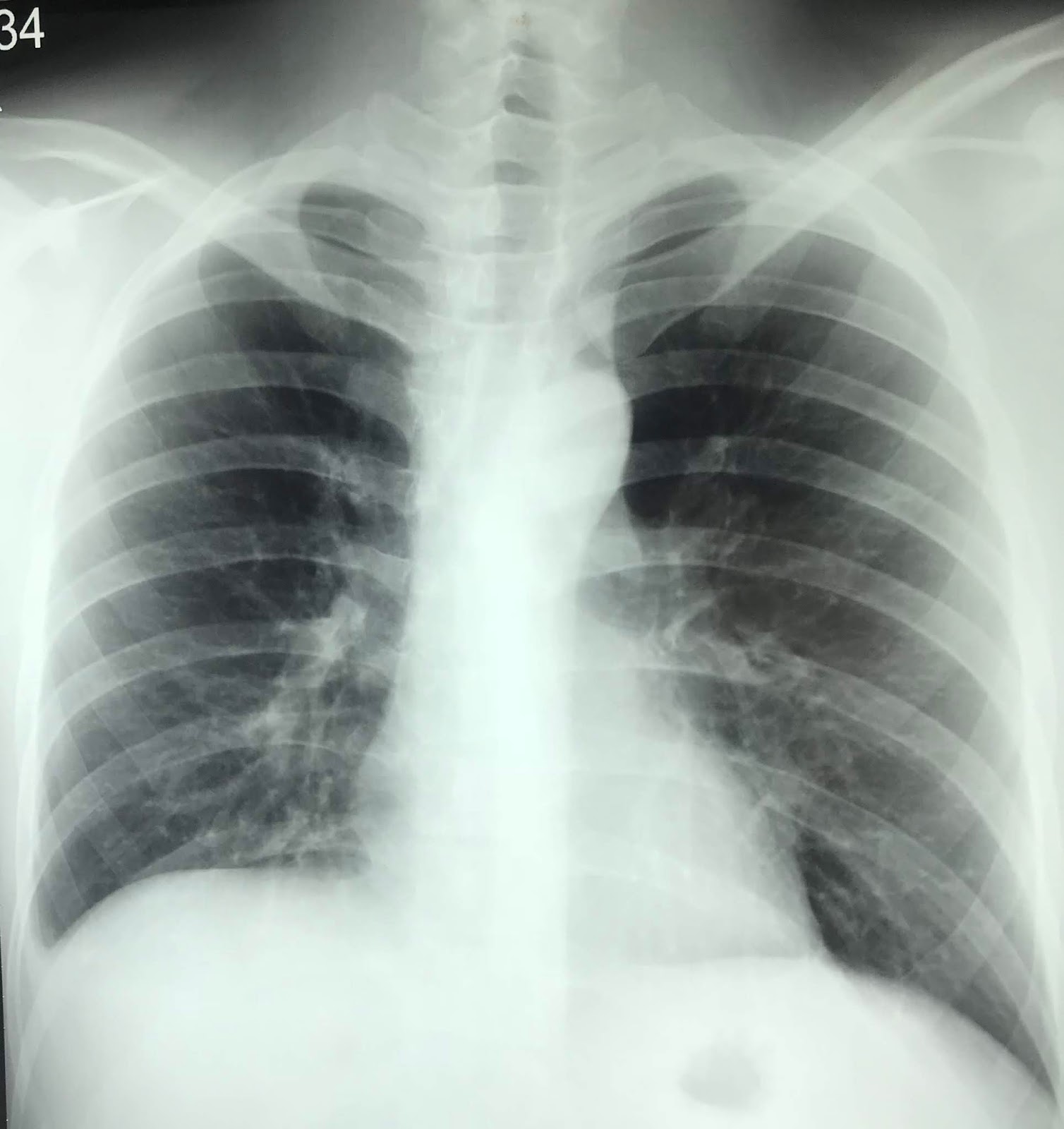
Woman 45 yo  with pain at left thorax.   
Chest X-rays detected one mass at left  lower lung.


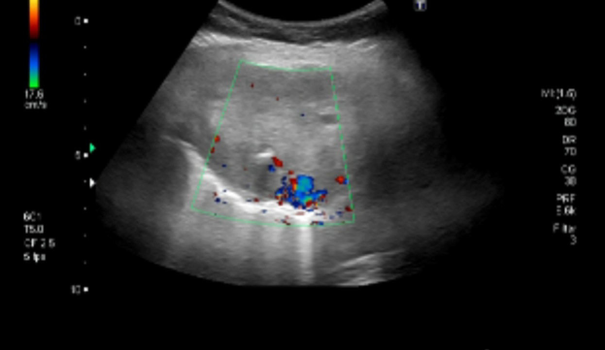
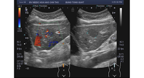
Ultrasound detected this mass from thorax wall.  US 1 :  solid mass  hypovascular.


US 2:  crossed section of this mass is round border freely with pleural space.


US 3 : longitudinal scan of this tumor is  hypovascular.


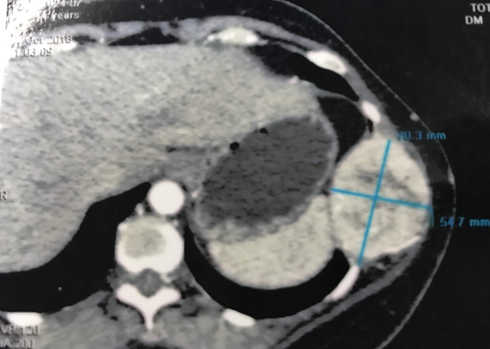
MSCT with CE of thorax,  this tumos is from 9th  rib, size 9cmx5 cm   ( CT 1  cross section,  C T 2   sagittal  section,   CT 3  3 D  view).


Biopsy of this mass is cavernous hemangioma.  
Operation for resection this tumor is done.
See tumor specimen.


Microscopic report is metastasis from thyroid cancer.

Review MSCT total body of this patient we see the thyroid tumor and liver focal which suspected metastasis from thyroid cancer.


Notes: This case is one of 18 thyroid cancer cases  and one of 4 cases bone metastasis published on VUD.

Wednesday 6 March 2019

CASE 540: URINARY BLADDER TUMOR, Dr PHAN THANH HAI, MEDIC MEDICAL CENTER, HCMC, VIETNAM.


MAN 32 YO  with HEMATURIA.  

ULTRASOUND of ABDOMEN.  
US 1 : LONGITUDINAL SCAN AT HYPOGASTRIC  REGION DETECTED  ONE CYSTIC MASS INTRA URINARY BLADDER WALL.  


US 2  : THIS MASS IS  FIXED TO ABDOMEN WALL .


US 3 : CROSSED-SECTION OF  CYSTIC MASS  IN URACHUS AREA.


CYSTO-ENDOSCOPY  DETECTED  ROUND TUMOR  MUCOSA OF UB INTACT.


MRI OF   URINARY BLADDER .
MRI 1, MRI 2  =  TUMOR 3 CM  INTRA UB WALL  CYSTIC 


MRI 3  DWI,   MRI 4 =  TUMOR IS  LOCATED AT URACHUS REMNANT.


OPERATION  FOR PARTIAL CYSTECTOMY.
OPE 1= AFTER  INCISION  THE TUMOR IS CYSTIC FILLED WITH MUCUS.
OPE 2 = FLUID CLEAR AND MUCUS GOES OUT.


MICROSCOPIC REPORT  is urachus cyst .


Thursday 28 February 2019

CASE 539: DENGUE FEVER and ASCITES, Dr PHAN THANH HAI, MEDIC MEDICAL CENTER, HCMC, VIETNAM.


MAN 25 YO WITH 3 DAYS  FEVER  AND EPIGASTRIC DISTENTION.
ULTRASOUND  FIRST :  
US 1: HEPATOMEGALY.  

US 2: GALL BLADDER WALL IS THICKENING 3 MM  NO STONE.

US 3:  FLUID IN MORRISON SPACE ( LIVER KIDNEY SPACE).

US 4: FLUID AROUND RIGHT KIDNEY.


US 5: IN PELVIS  BIG VOLUME OF ASCITES.

US 6: NORMAL SPLEEN WITH FLUID IN HILUS.


CHEST X-RAYS SHOWS  SMALL FLUID IN COSTO-PLEURAL SPACE.

BLOOD TESTS  SHOW   WBC HIGH, CRP HIGH  PLATTELETS  DROPPED TO 19K.          
ELISA  DENGUE FEVER TESTS  POSITIVE FOR IgG  AND IgM.


EMERGENCY ADMISSION IN INFECTIOUS TROPICAL HOSPITAL.

REFERENCES:
Role of ultrasound in Dengue hemorrhagic fever in Vietnam
http://www.vusta.vn/vi/news/Thong-tin-Su-kien-Thanh-tuu-KH-CN/Gia-tri-cua-sieu-am-bung-trong-sot-xuat-huyet-Dengue-21155.html
Ultrasound Findings in Dengue Hemorrhagic Fever

Monday 25 February 2019

CASE 538: SUBMERGED MEDIASTINUM GOITER TUMOR, Dr PHAN THANH HAI, MEDIC MEDICAL CENTER, HCMC, VIETNAM.


Woman 59 yo, for 10 years, underwent an operation of removing tumor at left lobe of thyroid.
Now she suffers from cough and  change of the speaking voice.
Chest X-Rays shows one mass at upper part of retrosternum.


Ultrasound  of the neck: 

US 1: crossed section of the right big thyroid lobe with tumor.


U S 2 : this tumor is prolonged from retrosternum. to mediastinum.


US 3: CDI with  hypovascular tumor.


US 4: longitudinal tumor scan.


Blood tests: normal thyroid function.

MSCT with CE:


CT 1, CT 2, CT 3                                                                                                                                                            Section  at level of neck  upper sternum  and middle  sternum   this tumor  from right lobe thyroid  calcification.


CT 4 : sagittal,  C T 5 : frontal section.


Isotopic Tech 99m  scan  made  diagnostic is thyroid tumor with cold nodule come from r ight lobe of thyroid gland.


Operation by the way sternotomy  removed this tumor well bordered,  central solid and cystic parts very highly calcified.


Conclusion:  Goiter tumor  submerged in mediastinum.

MICROSCOPIC REPORT IS  NODULAR GOITER.


Saturday 16 February 2019

CASE 537: GASTRIC LYMPHOMA, Dr PHAN THANH HAI, Dr LE THI THANH THAO, MEDIC MEDICAL CENTER, HCMC, VIETNAM.


Man 63 yo with gastric ulcer being treated for a  long time but not getting better. 
Gastroendoscopy  found a crater ulcer of antrium  but  in 2 times of biopsy only chronic inflamation.


Ultrasound scanning in epigastris  detected antrium thickening of the wall  and hypoechoic (US 1).   

US 2 : many lymph nodes around antrium.  


US 3: CDI, vascular supply for antrum.


US 4: longitudinal scanning of antrum  is  thickening of  the gastric wall.


MSCT scanning of abdomen.
CT 1= crossed section of   antrum with the mass.


CT 2 = scanning with position rotation of antrum showed stenosis.


Radiologist and sonologist diagnostics were gastric cancer.
Operation of gastrectomy.
Specimen 1= antrum tumors.  

Specimen 2 =
Opening of gastric lumen there wered 2 lesions at antrum and body of stomach.


Microscopic report with immuno-histo chemistry  staining is B cell lymphoma  of antrum and infiltrated lymph nodes.