Total Pageviews

Sunday 16 December 2018

CASE 529: CC HCC, Dr PHAN THANH HAI, Dr DUONG NGOC THANH, MEDIC MEDICAL CENTER, HCMC, VIETNAM.


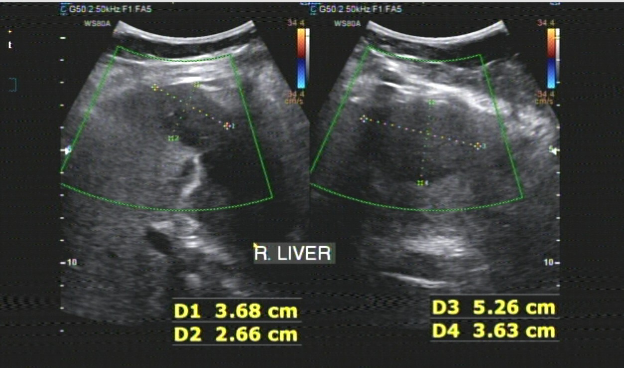
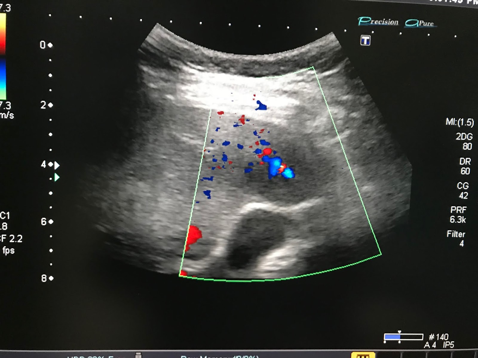
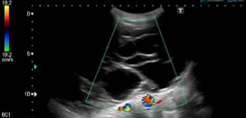
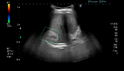
WOMAN 66 YO,   ULTRASOUND CHECK -UP DETECTED ONE MASS  5 CM AT R LIVER   HYPERECHOIC WELL- BORDERED  LOOKS LIKE  HEMANGIOMA (US 1, US 2, CENTRAL TUMOR NECROSIS).


BLOOD TESTS=  HBV AND HCV NON REACTIVE   WAKO TEST  STRONG POSITIVE.


MRI OF LIVER WITH GADOVIST , THIS TUMOR IS ENHANCED WITH GADO AND  STRUCTURE IS MORE FATTY TISSUE .  RADIOLOGIST REPORT IS AML LIVER ( MRI 1, MRI 2, MRI 3).


 CORE BIOPSY REPORT IS MORE FATTY TISSUE  WITH THE  SAME NUMBERS OF ABNORMAL CELL.

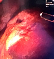
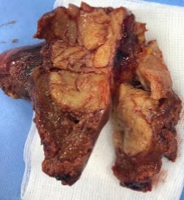
OPEN SURGERY RESECTION OF TUMOR  ( SEE MACRO  TUMOR WHICH IS  VERY DIFFERENT WITH LIVER TISSUE , WHITE  HARD   CENTRAL NECROSIS  WELL-BORDERD. MACRO 1, MACRO 2).


MICROSCOPIC REPORT IS  CLEAR CELL HCC   MORE 50%  CLEAR CELLS IN TUMOR.


Monday 10 December 2018

CASE 528: LIVER TUMOR, Dr PHAN THANH HAI, Dr JASMINE THANH XUAN, MEDIC MEDICAL CENTER, HCMC, VIETNAM.


Woman 69 yo with history of mastectomy due to breast cancer for 18 years. Now for a screening  she chose full body CT.
CT reported a liver mass   at segment 5 ,  size# 6cm  (CT 1 and  CT 2).


Ultrasound reviewed:

 US 1: Liver  picture scan in April 2018  is  normal

US 2 :   A hypoechoic mass near the gall bladder.


US 3: Vascular supply for this mass is central tumor.


Blood tests= Wako test normal,  CA 15-3  normal .

MRI of liver with primovist  made  diagnostic  by radiologist is metastasis to liver ( MRI 1,  2,  3, 4).


Ultrasound guided  core biopsy  report = fibrosis

Summary of this case:
 A  mass in liver # 6 cm  suspected metastasis from breast cancer had been
 treated 18 years before and waiting for operation.
Operation of robotic laparoscopy removed big tumor and gall bladder.

Microscopic report is metastasis from suspected breast cancer.


MICROSCOPIC REPORT WITH IMMUNO-HISTOCHEMISTRY IS  METASTASIS MAY BE FROM BREAST CANCER  BEINGTREATED 18 YEARS AGO.


Reference   TNM  AND M DELAYED.


Monday 3 December 2018

CASE 527: SPINE METASTASES, Dr PHAN THANH HAI, MEDIC MEDICAL CENTER, HCMC, VIETNAM.


WOMAN 50 YO  3 YEARS AGO BEING TREATED  AT CANCER CENTER BY NPC  RADIOTHERPY AND RADICAL NECK DISSECTION. BUT NOW PAIN AT DORSAL  AREA.
ULTRASOUND OF PARAVERTEBRAL  AREA DETECTED  ONE MASS HYPOECHOIC AT THE LEFT LATERAL OF THE D10 SPINE.
 US 1, US 2  LONGITUDINAL SCAN,  US 3  CDI  NO MORE COLOR DOPPLER SIGNAL.


MRI  SPINE    DETECTED ABNORMAL D10  ( MRI1)   MRI 2  FOCUS  D10, MRI 3   CROSSED SECTION  D10,  MRI 4  TUMOR  T2   CALCIFICATION INTRATUMOR , MRI5  FRONTAL VIEW  NEAR  KIDNEY  UPPER POLE.


CORE BIOPSY  REPORT  IS  METASTASIS   BY  UNDIFFENTIATED CELL TUMOR .


Wednesday 28 November 2018

CASE 526: BIG OVARIAN CYSTIC TUMOR, Dr PHAN THANH HAI, Dr TRAM THI TU HUONG, MEDIC MEDICAL CENTER, HCMC, VIETNAM


FEMALE PATIENT 17YO,   SINGLE,  IRREGULAR MENTRUAL CYCLE AND  ABDOMEN DISTENTION.
ULTRASOUND  DETECTED  BIG CYSTIC TUMOR FROM PELVIS TO EPIGATRIC AREA.
US 1 : LONGITUDINAL SCAN OF ABDOMEN AT MIDDLE LINE.


US 2 : AT PELVIS.

 US 3 : CROSSED-SECTION AT ILIAC ARTERY DIVISION.  


US 4 : SMALL UTERUS AND ASCITES AROUND.


MSCT  WITH CE.
CT1 : CROSSED-SECTION OF ABDOMEN AT KIDNEY LEVEL.


CT2 : CROSSED- SECTION OF TUMOR.


CT3 : FRONTAL VIEW  


BLOOD TEST : ROMA TEST NEGATIF  
SUMMARY =IT IS BIG OVARY CYSTIC TUMOR, SIZE 50CM,  MULTISEPTATION.  IOTA CLASSIFICATION B4.
OPERATION REMOVED RIGH BIG OVARY TUMOR CASE.


IOTA Classification


MICROSCOPIC RESULT IS MUCINOUS CYSTADENOMA BORDERLINE MALIGNANCY of OVARY.


Sunday 18 November 2018

CASE 525: LIVER ABSCESS, Dr PHAN THANH HẢI, Dr NGUYỄN THÀNH ĐĂNG, MEDIC MEDICAL CENTER, HCMC, VIETNAM


Male  62yo, with history of removing biliary stones in liver for 10 years. 
Ultrasound in 5 times shows biliary stones intraliver, aerobilia and right liver cyst from liver cirrhosis and splenomegaly patient. Blood tests= WBC:19,800g/L, hs CRP=177.2H. AFP=19.8ng/mL.


He came back Medic for reexamination as pain at RUQ and slight fever in Feb 2018.
Ultrasound suspected a liver tumor  with biliary stones and cirrhosis and splenomegaly.


But MRI thought about a new liver abscess with biliary stones of cirrhosis patient.


He entered CR hospital and  the results of ultrasound and CT  were liver tumor and aerobilia. With the diagnosis of CR hospital on liver tumor necrosis and aerobilia he felt no pain and nor fever after using of prescribed medicine drugs in 17 days.


He came back Medic  2 months later. 
Ultrasound detected no liver tumor, only biliary stones and aerobilia. 


MRI revealed no liver tumor, and in reviewing,  MRI noted a sign of head of cauliflower that means a pyogenic abscess  appearing of peripheral lobulated increasing signal intensity on MRI.

SUMMARY: Ultrasound was in mistake as no pay attention of clinical signs. A tumor necrosis could not be in concordance with infectious syndrome. And patients remained quite well because of resolving of liver abscess.

 REFERENCE: