Girl 10yo with
epigastric pain.
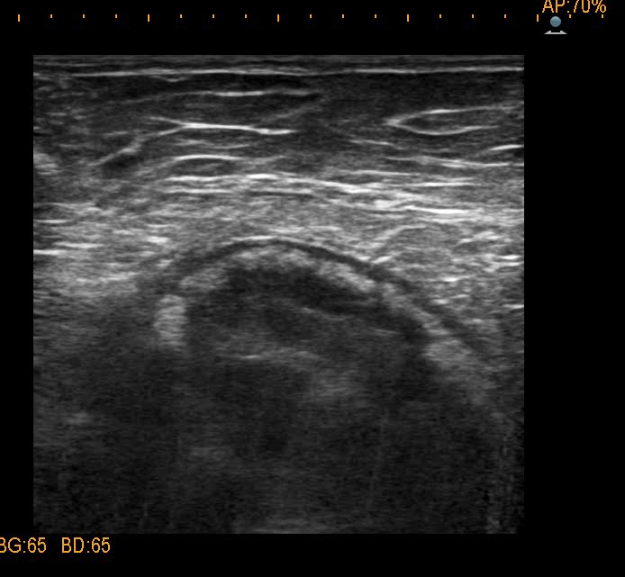
Ultrasound of abdomen
detected one mass of 8cm at the body of pancreas, cystic
structure, well bordered ( US 1, US 2, US 3), no lymph node
around.


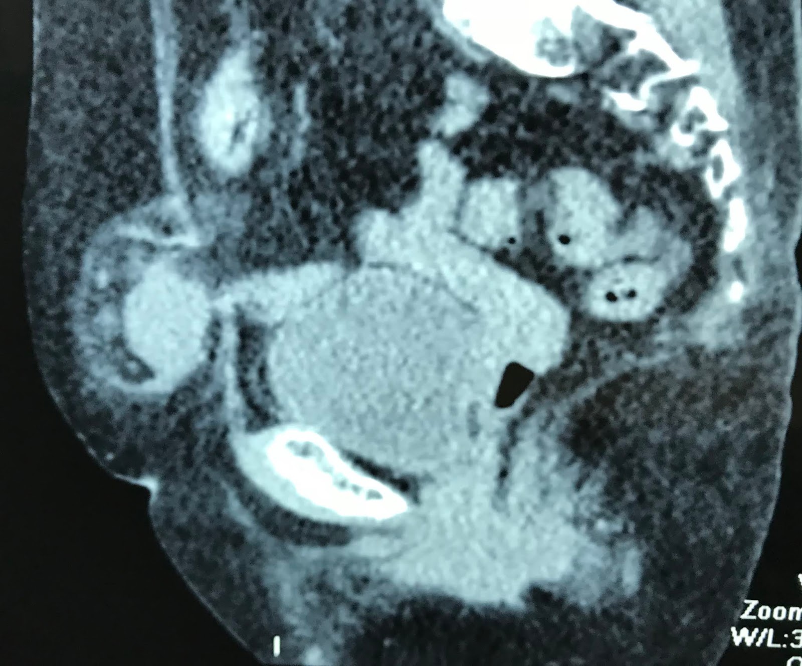
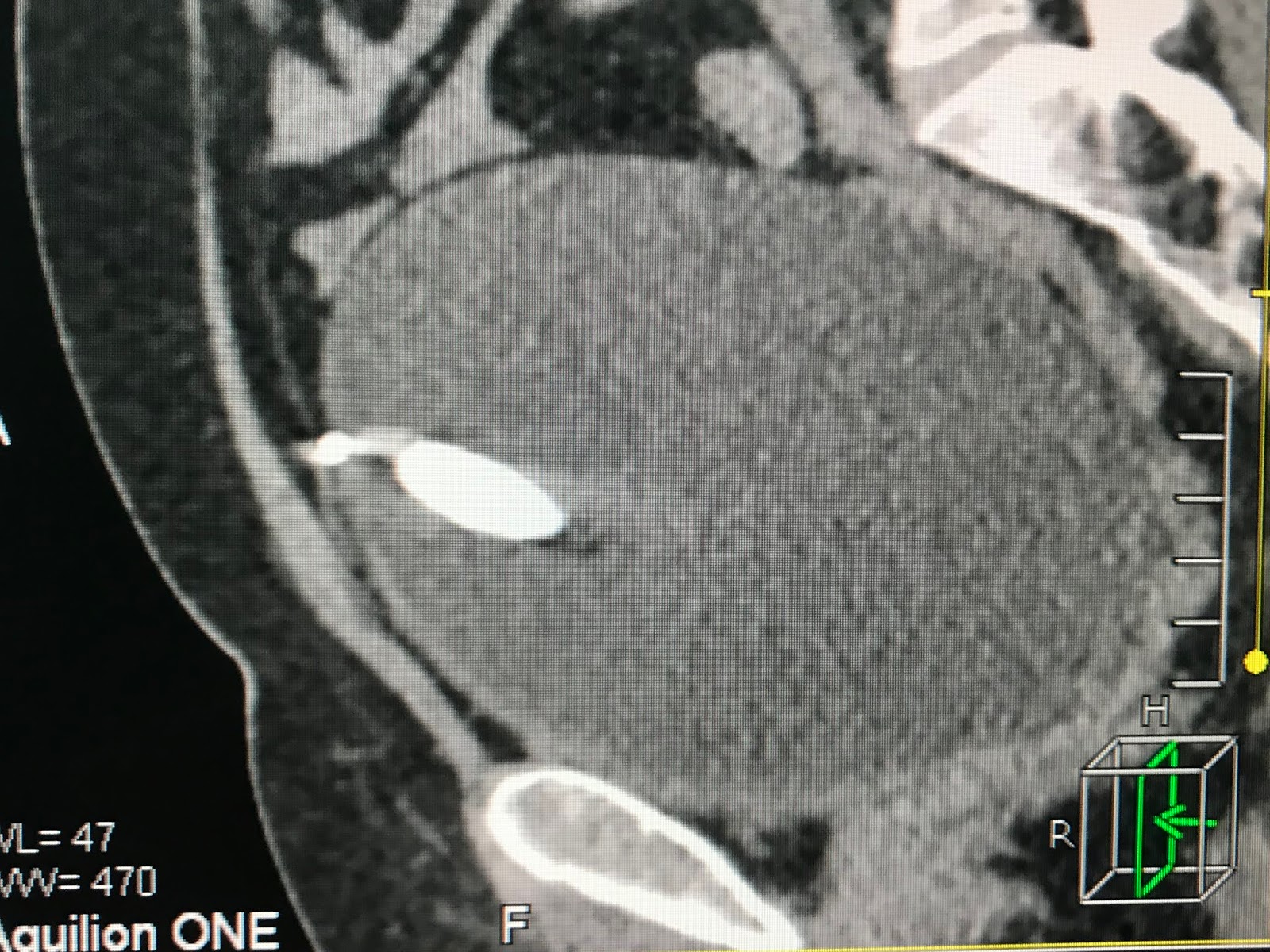
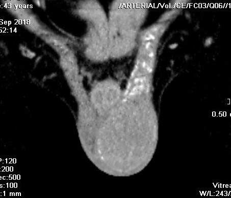
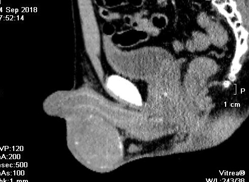
MSCT with CE=
this mass is from the tail of pancreas, inhomogeneous
structure (CT1 CT 2, CT 3).


Blood tests are
normal.
Operation for
resection of this tumor [see macro].

CT SHOWS THIS TUMOR A SOLID TUMOR BUT ULTRASOUND SCAN FINDING IS CYSTIC.
Summary: Girl 10
yo with big mas at the pancreas, structure is mixed solid and
cystic, the most common is solid pseudocystic papillary
tumor.
Histology result is pancreas pseudopapillary neoplasia.

REFERENCE:PDF.