Woman 42 yo with
right breast hard mass,clinical examination of asymetric thorax (photo).

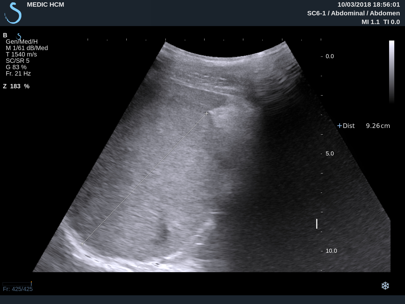
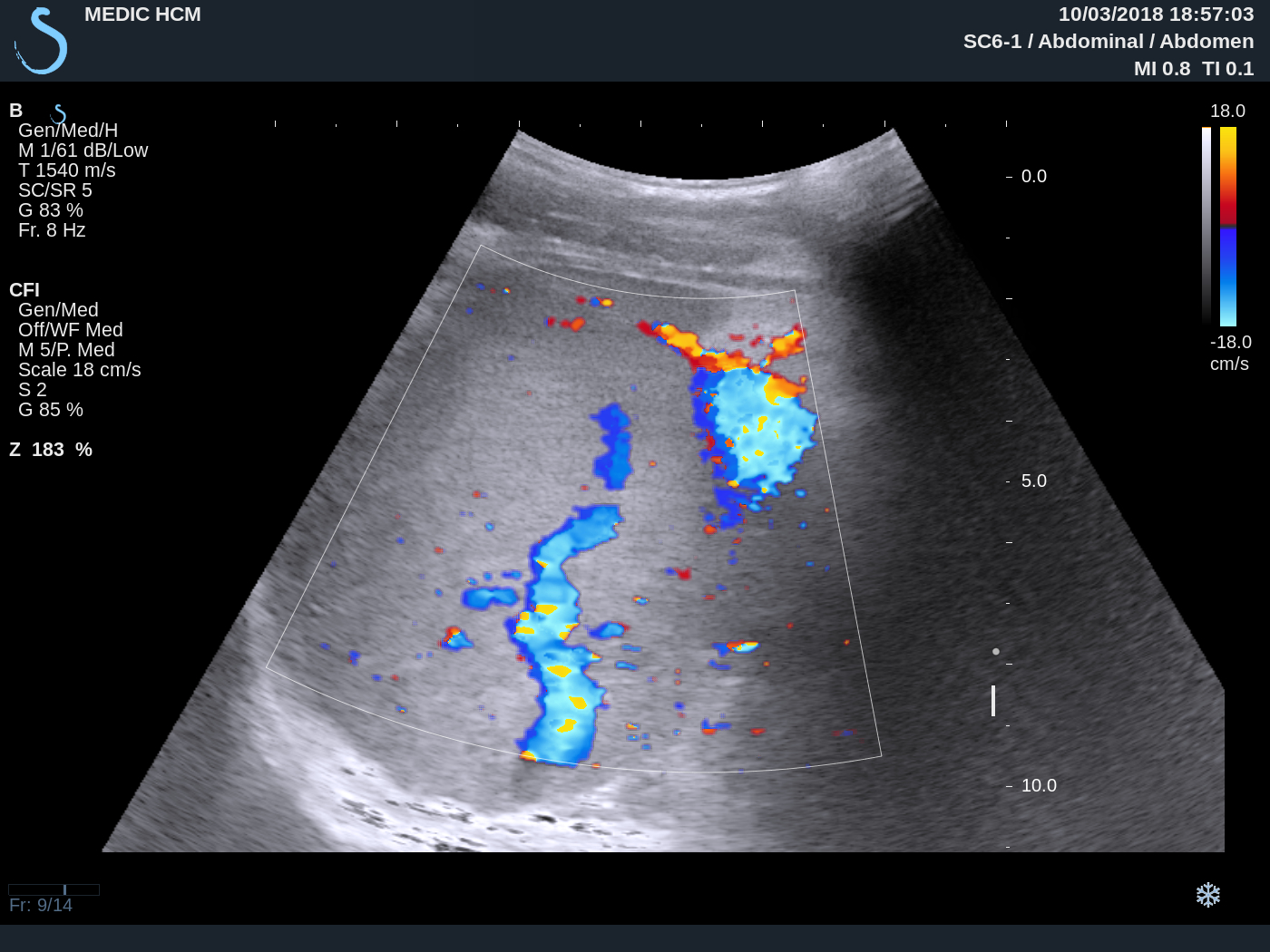
Ultrasound of
right breast detected one 5 cm mass, multilobular, hypoechoic with blood
supply arround this tumor ( US 1, US 2 ), US 3 detected axillary
lymph node, US 4 in comparison of right to left chest wall shows absence
of right major and minor pectoralis muscles.


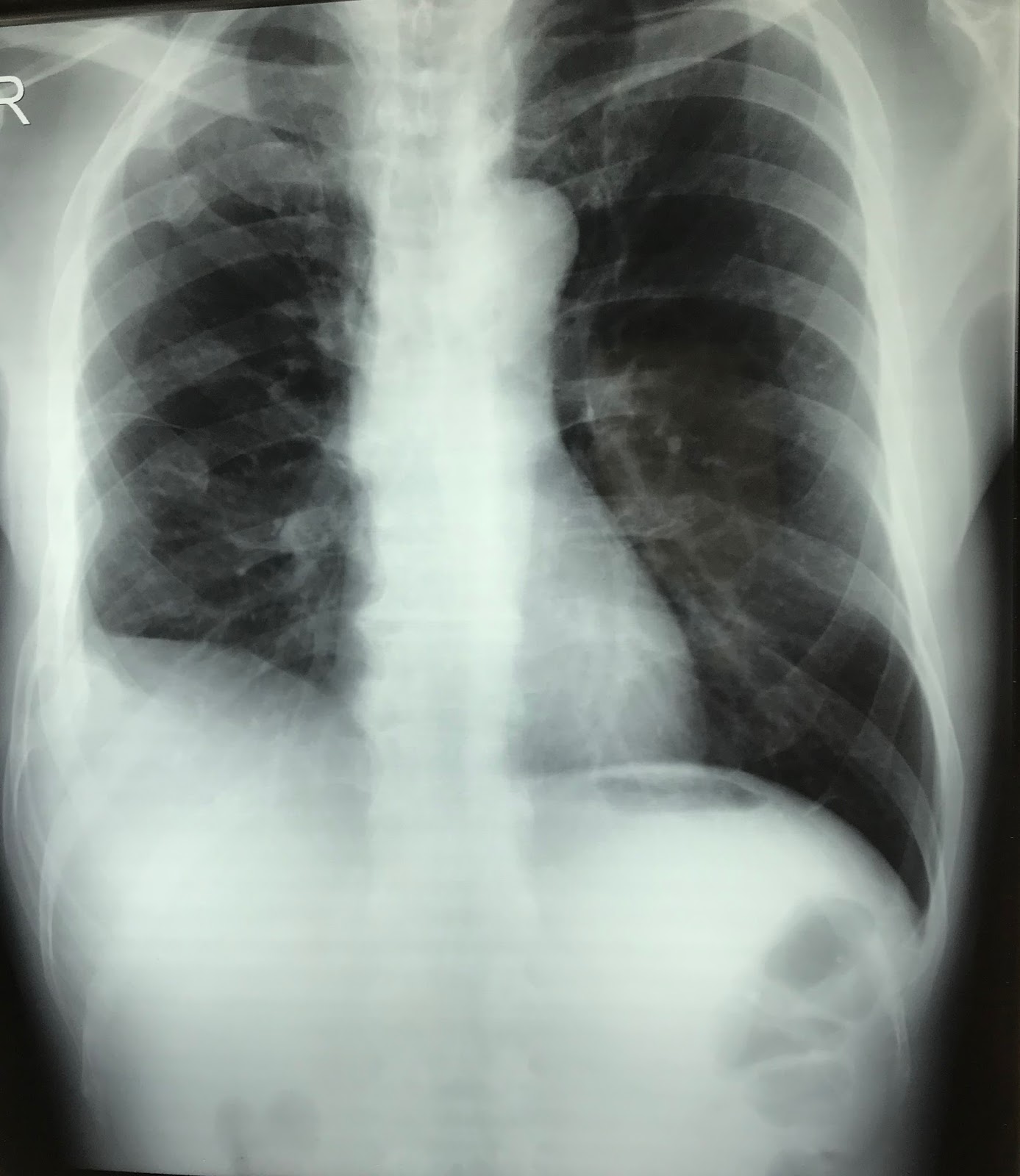
Chest X-Ray :
clear right lung in comparison to left lung due to right chest wall
muscle defect.

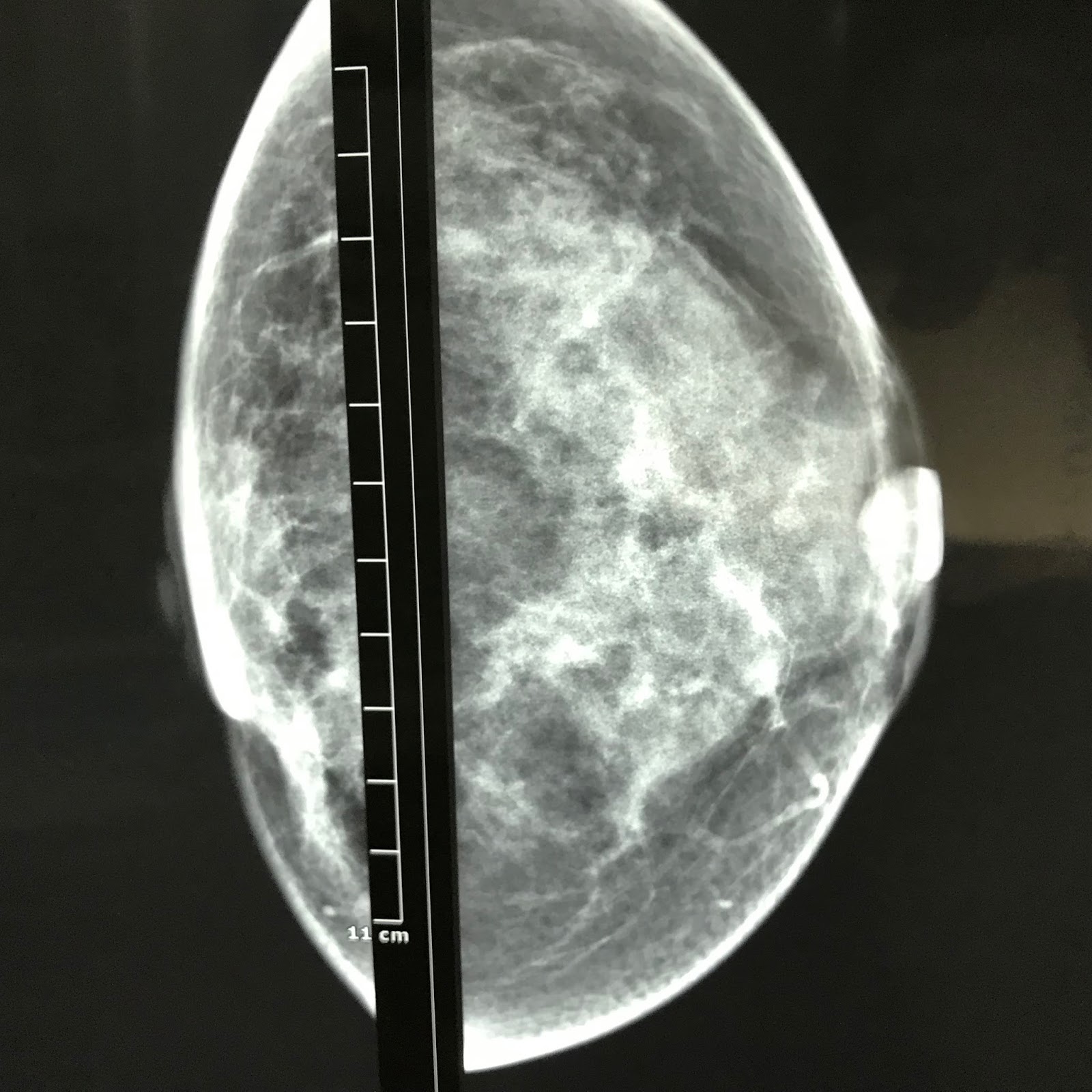
Mammography
diagnosis is breast tumor with Bi-Rads 4 T2N1Mx.

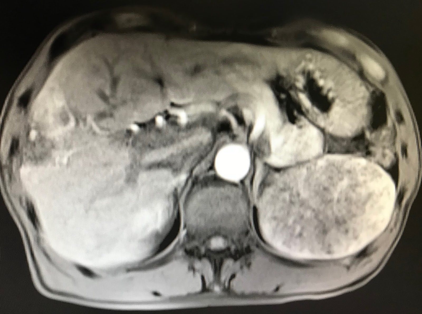
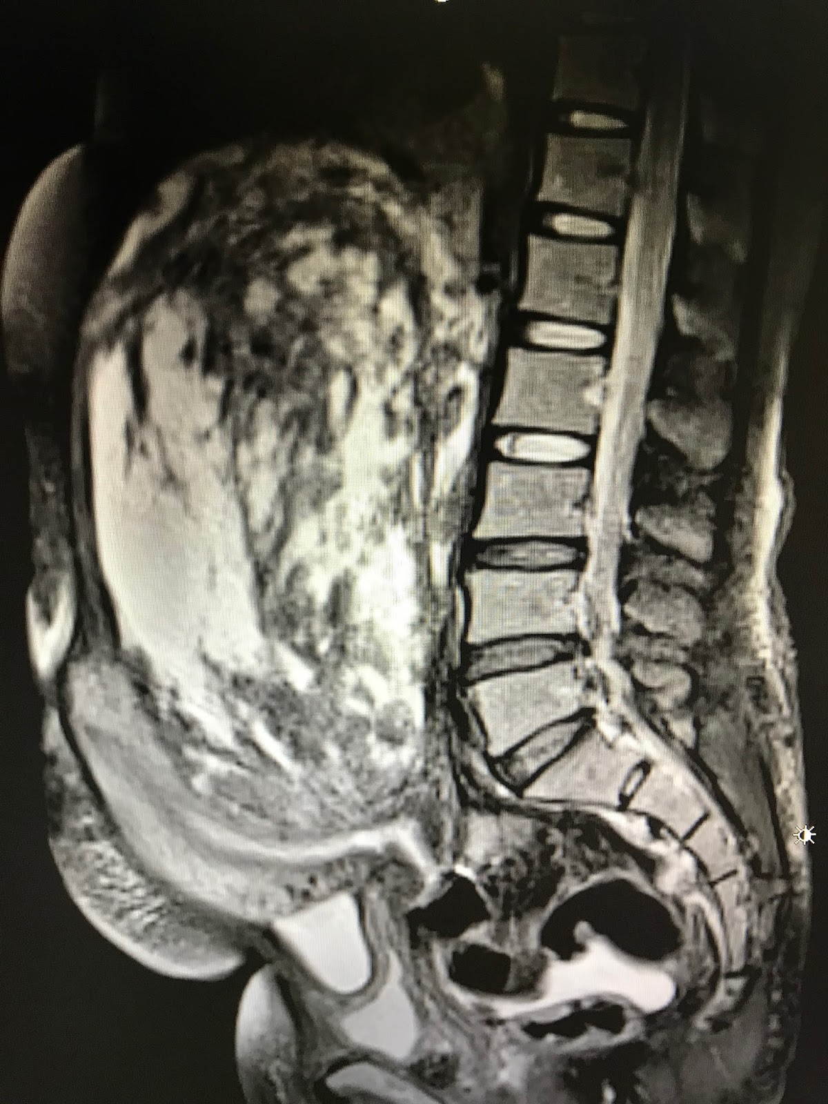
MRI made
diagnosis of right breast tumor with pectoralis
muscle defect of chest wall: it is Poland’s syndrome.

FNAC of this tumor with cytological report of adenocarcinoma of breast tumor.