Total Pageviews

Thursday 1 February 2018

CASE 475: INCIDENTAL THYROID CARCINOMA ON CT, Dr PHAN THANH HẢI, MEDIC MEDICAL CENTER, HCMC, VIETNAM.


Man 55 yo for check- up  MSCT  total body and blood tests.

Radiologist report  detected one mass # 1.6 cm at right lobe  of thyroid gland,  with  HU 91 UI  in comparison to  the thyroid tissue background =121UI ( CT1  CT2  CT3).


Blood test is normal thyroid function.

Ultrasound scanning is second look:

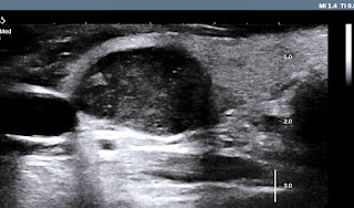
US 1:  crossed-section  of the hypoechoic  focal  lesion, well-bordered #  1.5 cm.
US 2:  longitudinal scanning of  the mass is 1. 7 cm, hypoechoic pattern,  with 
US 3:  CDI = hypovascular  mass,  no lymph node in the neck.


This mass is  in TI-RADS 3, need  FNAC.
FNAC report suspected  papillary carcinoma ( PTC).


Operation is done for subtotal thyroidectomy (see macro 1,2).


POST OP  PATHOLOGY REPORT IT IS PTC [PAPILLARY THYROID CARCINOMA].


REFERENCE:


Friday 26 January 2018

CASE 474: LEFT LIVER LOBE TUMOR, Dr PHAN THANH HẢI, MEDIC MEDICAL CENTER, HCMC, VIETNAM


Man  52 yo  with HBV chronic hepatitis ,    follow up  every 6 months  and negative AFP, HBV -DNA . But  ultrasound   detected  a big tumor # 6 cm in diameter in left lobe of liver.
US  1:  transverse scanning of  this tumor is well bordered,   hypoechoic pattern.
US 2:  longitudinal scanning  at tumor site.
US 3:  color Doppler=  vascular supply to tumor is from left  liver.


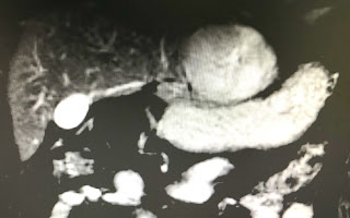
MRI  with Primovist  uptaked  and washed out  as a HCC in liver ( MRI 1, 2, 3, 4).


Blood test  Wako=  only DCP  raised =91 UI.


Summary:  in case of  HBV chronic hepatitis,, ultrasound detected  a big tumor in liver, Wako test only raised DCP.

OPERATION   PER OP  VIEW BY ENDOSCOPY THE TUMOR AT LEFT LOBE of LIVER ANTERIOR AND POSTERIOR and MACRO SPECIMEN of  TUMOR.


MICROSCOPIC IS WELL DIFFERENTIATED HCC.


DISCUSSION: WHY IN THIS CASE   WAKO TEST IS  NOT SUITABLE ?  WHICH VALOUR of WAKO TEST COULD BE  PPV FOR HCC ?

 Wako test post op  4 days after operation  = AFP:1.3 ng/mL;  L3: 0.5%;  DCP: 55 mAU/mL.

This 52 yo male  patient with chronic HBV but  AFP is lower than cut of value screening and no  ultrasound  screening  before  operation.   Wako test is  only DCP  rising  to 92 mAU/mL. Reference  ( publication April 12,2016 http:// doi.org/10.1371/journal.pone.0153227  : Diagnostic evaluation of DCP versus AFP  for Hepatitis Bvirus related  HCC   in China):

1-  30-40%  HCC in CLD with  AFP normal serum level.
2-  non HCC patients  have  15-58%  AFP  rise  over  cutt of value  20ng/mL.
3-  in CLD having cirrhosis  AFP rises  11-47%  but non HCC.
4-  DCP  rises level that correlated with size tumor and advanced progress.
5 - DCP drops very fast after surgery, and  rises  early in recurrent HCC.

Conclusion : AFP only not sensitive for screening  HCC. So DCP in Wako test is the choice for routine screening  HCC and monitoring after treatment.

BLOOD TEST  WAKO  AGAIN    RESULT IS  AFP: 1,5;   L3 : 0,5;   DCP DROP TO 38mUI/mL


CONCLUSION  2:

DCP IS VERY SENSITIVE FOR DETECTION of HCC AND FOLLOW UP POST OP.

Tuesday 23 January 2018

CASE 473: SCIATIC NERVE TUMOR, Dr PHAN THANH HẢI, MEDIC MEDICAL CENTER, HCMC, VIETNAM.


Woman 59yo  detected  one mass  at right buttock that is in slow growth and no painfull.
Ultrasound scan
US 1= longitudinal scan of  this mass with size 6.7 cm,  ovoid, hypoechoic  in gluteus maximus muscle   near sciatic nerve  like a size of a mango.


US 2 = crossed section of this tumor is well bordered and hypovascular pattern. 

US 3 = elasoscanning of  this mass is inhomogeneous  structure.


MRI scan


MRI 1   crossed section   this mass is   well bordered  in a muscle   
MRI 2   frontal view this mas is  bordered of right sciatic nerve.
MRI 3   relation of this mass and right sciatic nerve.


Core  biopsy is done and histology report is   neuroma.


Friday 19 January 2018

CASE 472: INCIDENTAL BREAST TUMOR , Dr PHAN THANH HẢI, MEDIC MEDICAL CENTER, HCMC, VIETNAM


WOMAN 58YO   FOR SCREENING  TO DO WHOLE BODY MSCT. 
RADIOLOGIST REPORTED ONE MASS AT  RIGHT  BREAST  SIZE# 1.3 CM.(  CT1  FRONTAL VIEW, CT2  AXILLARY AREA).


ULTRASOUND POST CT VERIFIED THIS MASS  WITH  SIZE  # 2 CM   VERY STRONG  SHADOWING, AND HYPERVASCULAR.


US1,  US2  ELASTO  IS  31.9 kPa .
US3 , AXILLARY NODE    SIZE 1.5CM WITH HILUS 
US4 ,  CDI  = VASCULAR  HILUS DEFORMATION.


CORE BIOPSY  RESULT IS  BREAST CANCER, STAGE T2 N2.

OPERATION  for MASTECTOMY AND   AXILLARY   LYMPHADENECTOMY. SPECIMEN  TUMOR IS SMALL SCLEROSIS   MANY LYMPH NODES.


POST OP  HISTOLOGY REPORTED   THIS CANCER  METASTATIZES TO AXILLARY LYMPH NODES.

REFERENCE:


Sunday 14 January 2018

CASE 471: MESENTERIC LYMPHOMA, Dr PHAN THANH HẢI, MEDIC MEDICAL CENTER, HCMC, VIETNAM.


Man 70yo  with  epigastric pain. Ultrasound of abdomen detected  one hypoechoic mass like a cyst # 4.9cm near SMA.
US 1=   longitudinal scan at epigastric area.

US 2 = CDI:   this mass  is near SMA.

US  3 = crossed scanning= this mass  is placed over the head of pancreas in relation  with  celiac artery and vein.

US 4=  some  small lymph nodes near the big anesthetic mass.


MSCT CE=
CT  1 : crossed section  this mass  over the artery and vein.


CT 2:  the pancreas is normal.

   
CT 3:  frontal section of this intramesenteric mass near  SMA and vein   with some  lymph nodes in mesentery.


Endoscopy gastric and colon are normal, blood test and all cancer markers are normal 

Laparotomy for  biopsy= many nodes in mesentery.
Microscopic with histobiochemistry  is  follicular B CELL LYMPHOMA.


SUMMARY :  MANY LYMPH NODES IN MESENTERY  WITH TYPICAL APPEARANCE HYPOECHOIC PATTERN LOOKED LIKE A CYST,THE MOST COMMON IS LYMPHOMA.

Reference :    MEDIC ULTRASOUND case 452 : Castleman disease.