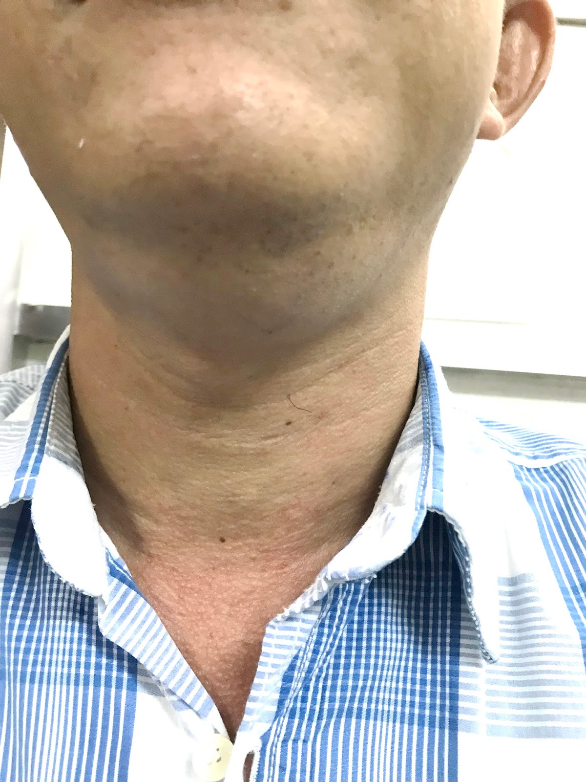
Man 41 yo, 5 years ago detected
one mass at submandibular region, slow growth. Clinical palpation no pain, soft, and without trouble of
eating.
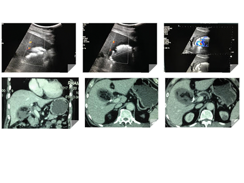
Ultrasound:
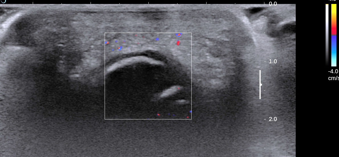
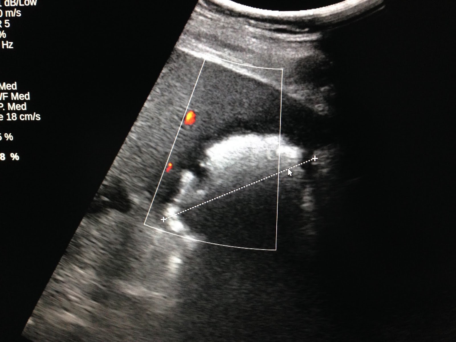
US 1= longitudinal scan with
curve probe 3.5 MHz : ovoid mass clear border, hypoechoic with
no vascular signals inside.

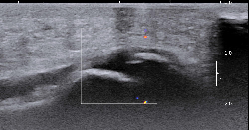
US 2 = scanning with linear probe 12MHz=
inhomogeneous structure with many
black spots, size # 0.5 cm.
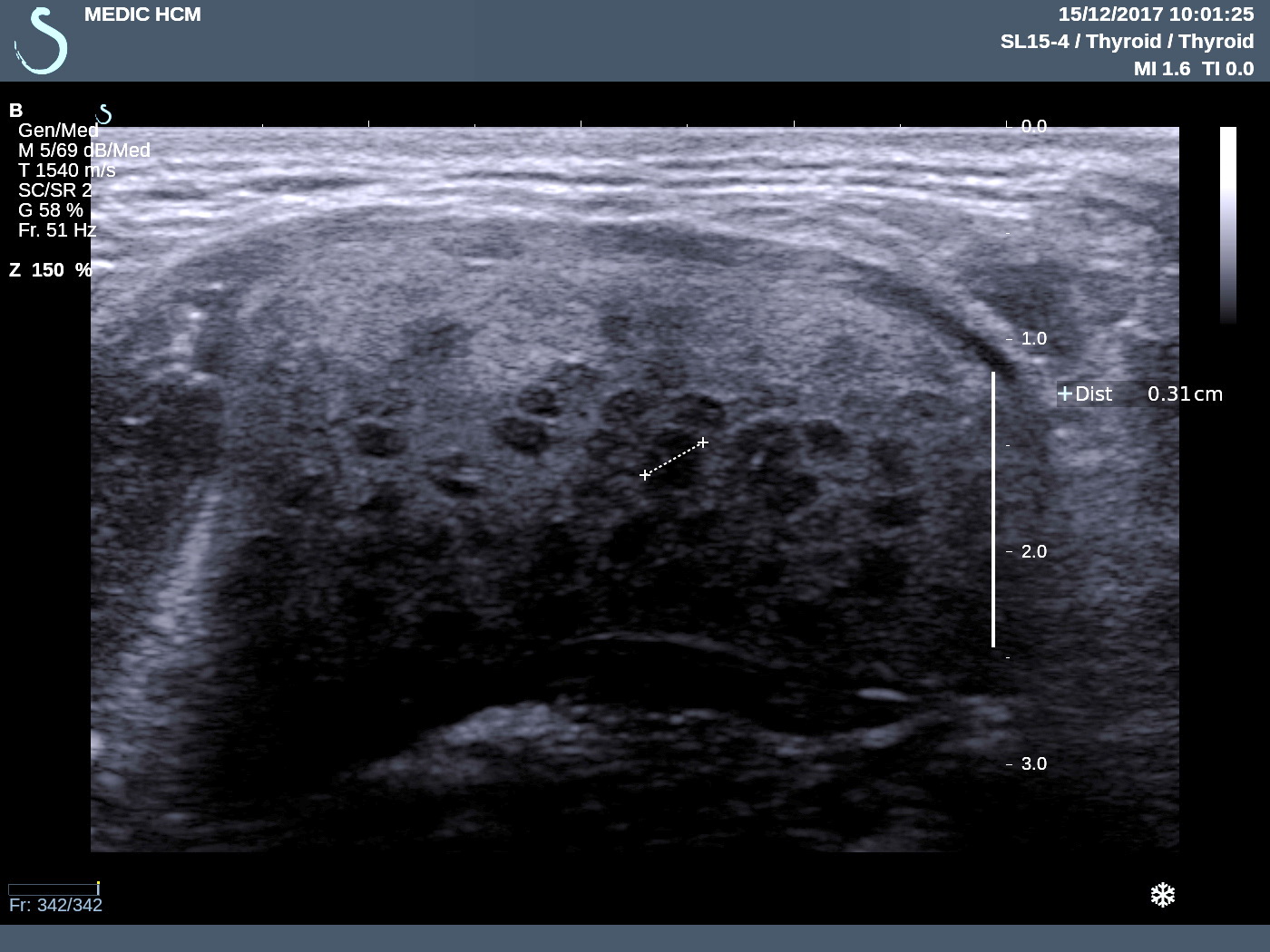
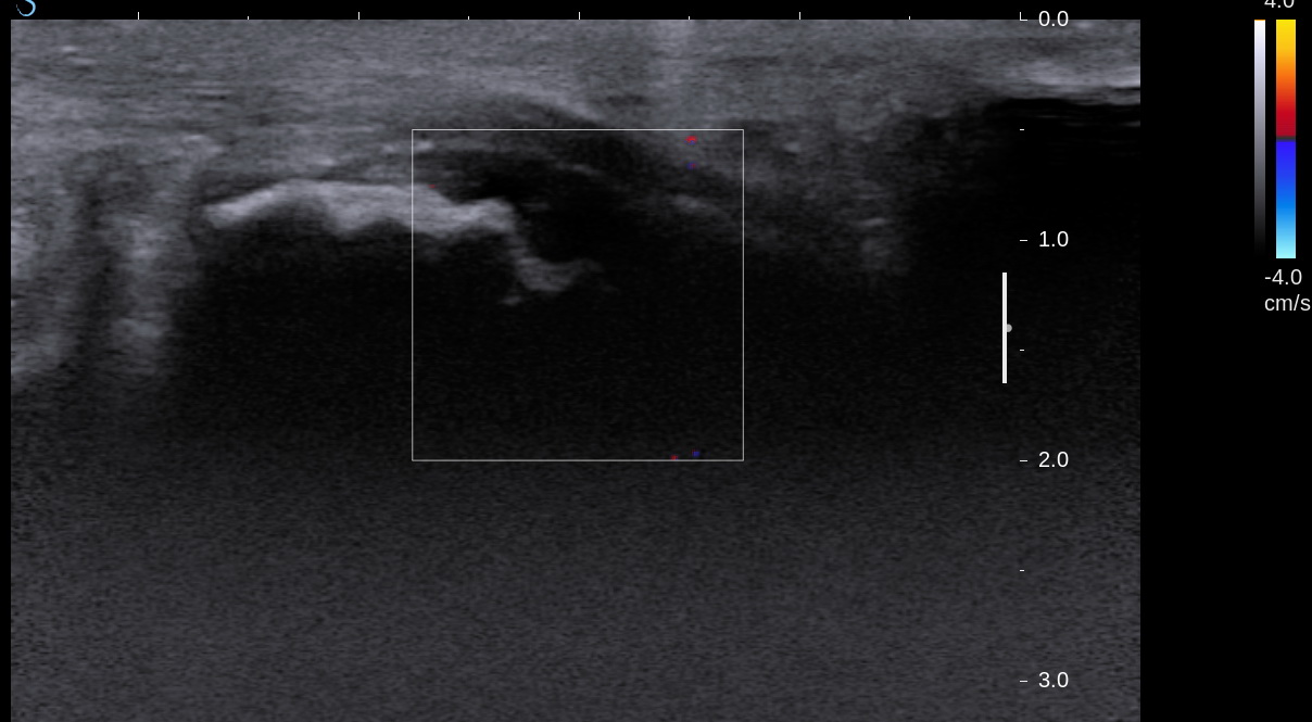
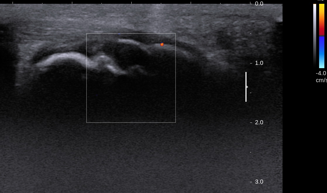
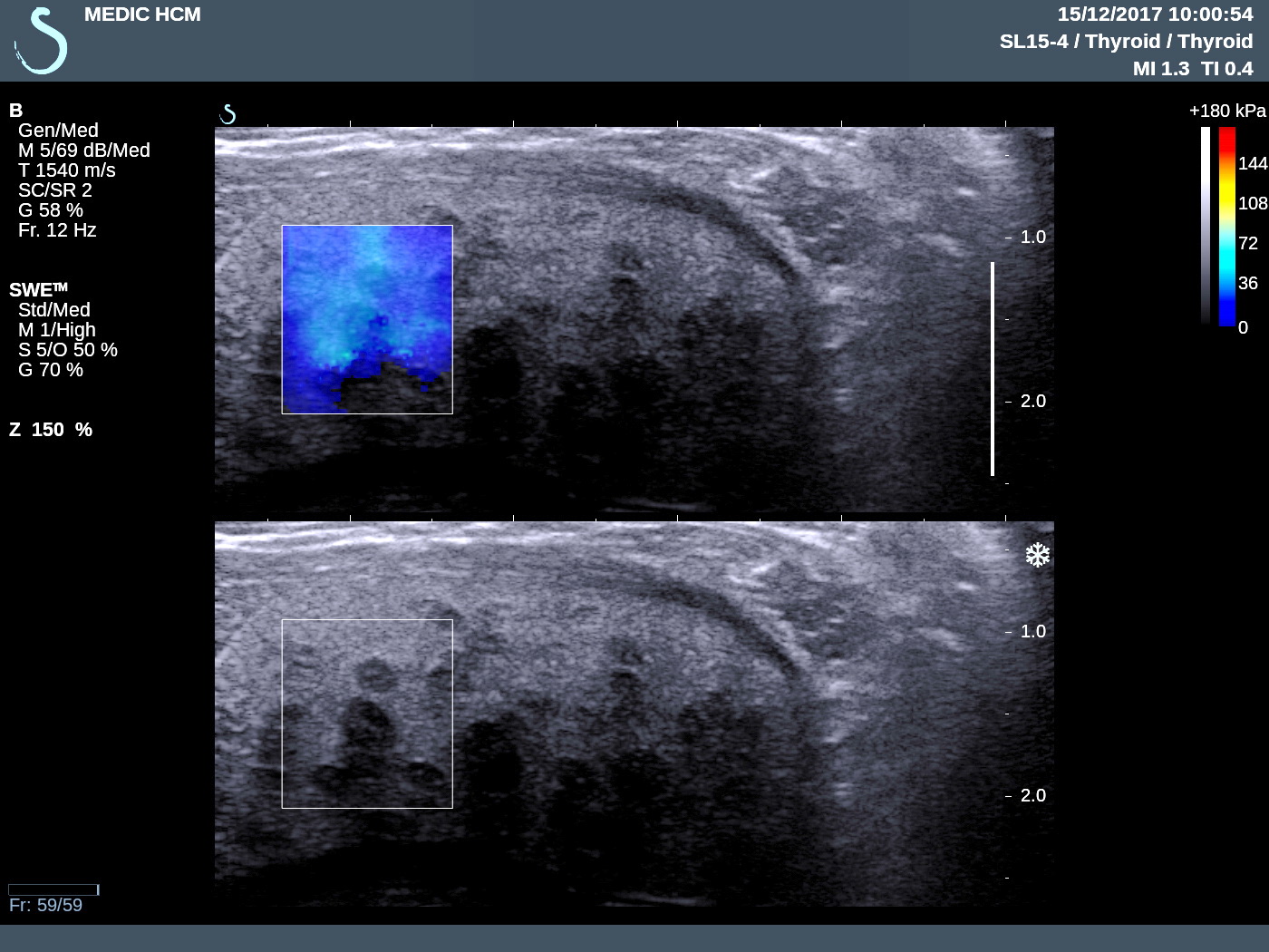
US 3 = elastoscanning of this mass disclosed
a cyst with many spots hardening; like pomegranate fruit.
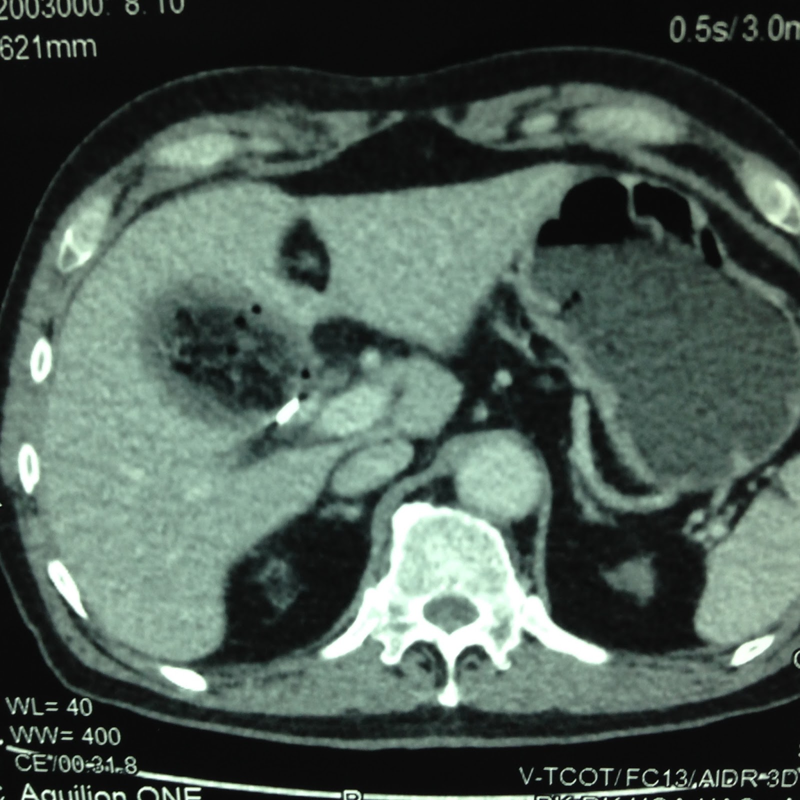
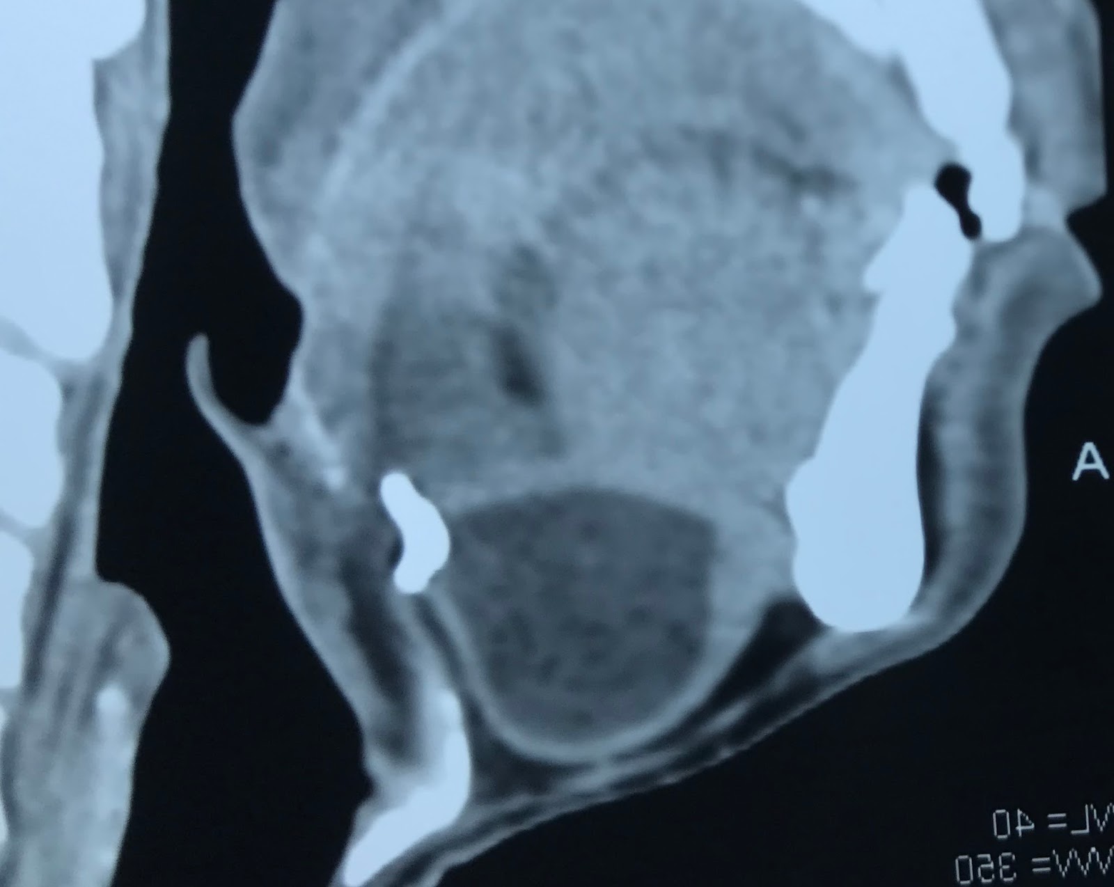
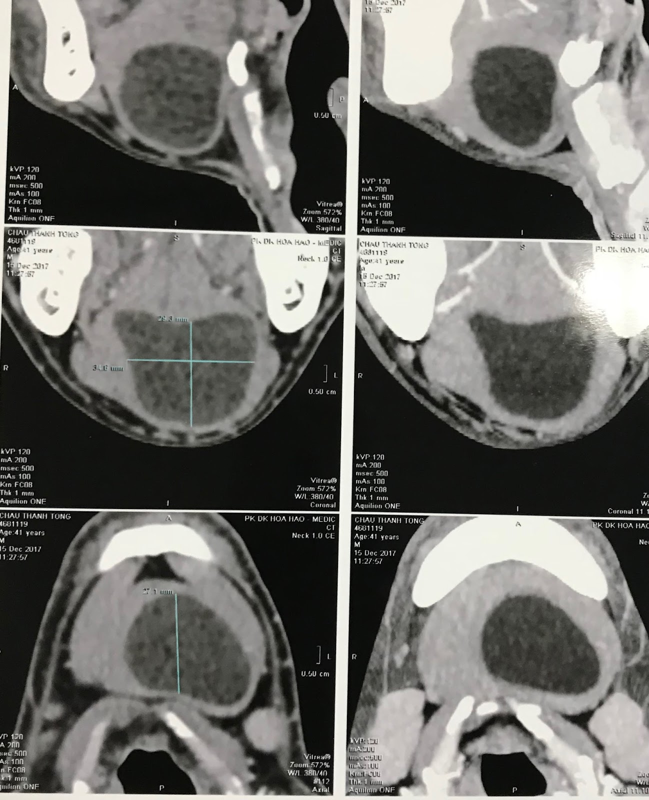
CT scan with CE= it is a cyst,
well bordered, .CT1, 2 , 3 with 3 sections of this mass,
radiologist said teratoma.

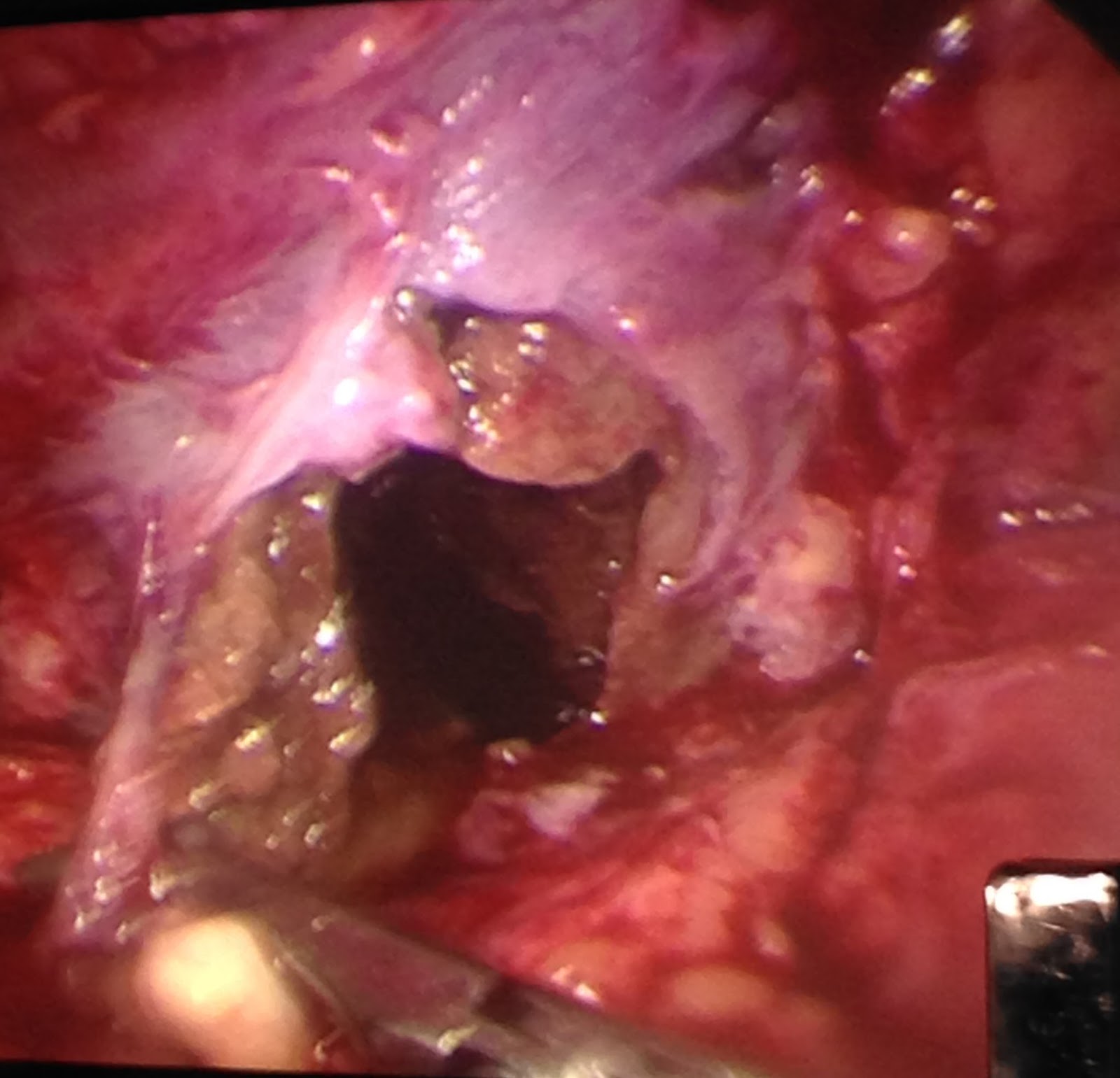
Operation for remove this cyst
with content like yellow milk typical of sebaceous
cyst (epidermoid cyst).

MICROSCOPIC REPORT IS EPIDERMOID CYST, BENIGN TUMOR.

REFERENCE: IT IS RICE BODIES.