Total Pageviews

Sunday 10 September 2017

CASE 451: CARDIAC MYXOMA, Dr PHAN THANH HẢI Dr NGUYỄN TUẤN VŨ, MEDIC MEDICAL CENTER, HCMC, VIETNAM.


Woman 33 yo, with  dypsnea progressing for one year but being normal chest X-Rays , EKG and abdomen ultrasound.   

  
Cardiac ultrasound  detected  one mass 5 cm intra left atrium  covering  near all cavity space ( US 1, US 2, US 3). 


And  echocardiologist says  cardiac myxoma.
CTA  of cardiology  non CE showed  this round mass  related to  left atrium, and radiologist confirmed  myxoma in left atrium.


Emergency heart operation was done.
OPERATION  REMOVED BIG TUMOR 5 CM  WITH  STRUCTURE LIKE  MYXOMA.


MICROSCOPIC REPORT IS MYXOMA.

Reference:


Saturday 2 September 2017

CASE 450: PID with PELVIC MASS, Dr PHAN THANH HẢI, MEDIC MEDICAL CENTER, HCMC, VIETNAM

Woman  34 yo with  onset 2 weeks ago,  fever  and pain at pelvic region.
Ultrasound  first at an obgyn hospital  says  ovary cyst or endometriosis. But medical  management  with antibiotis failed in clearing fever.


(US 1: scanning  after 2wks,  uterus and cystic mass # 10cm with very thickening wall; US 2 : on CDI;  US 3:  intracystic mass detecting one structure like a bridge: US 4:  ascites intra RLAQ;  US 5:  this mass was scanned with linear 12 MHz probe. 
MRI of pelvis with gado=   this cystic mass is  in left ovary  with  the wall very thick, and  black spot intra mass  unknown  original,   but radiologist  suspected an ovary cancer.


Blood tests=   WBC  12k with 70% neutron,  Plt= 515,  CA 125  rising  100 UI.


 18 DAY FEVER AGO CANNOT STOP BY ANTIBIOTICS.  OPERATION BY LAPAROENDOSCOPY WITH DIAGNOSIS  OF P I D.
Picture OPE1: LEFT OVARY IS VERY BIG MASS AND FALLOPEAN TUBE IS BIG ALSO.  


Picture OPE 2: INCISION OF THE WALL OF CYSTIC MASS SHOWING VERY THICK AND an AMOUNT of PUS  GOES OUT.


Picture OP 3: VASCULAR THROMBOSIS INTRACYSTIC MASS.


BACTERIOLOGY  REPORT  of THE PUS from STREPTOCOCCUS  and  MICROSCOPIC REPORT of THE WALL of CYST IS  ABSCESS WALL.


Reference:

Wednesday 30 August 2017

CASE 449: LIVER TUMOR, Dr PHAN THANH HẢI, MEDIC MEDICAL CENTER, HCMC, VIETNAM


Man 77yo, in  general check up  by ultrasound  detected  one round mass 5 cm in  segment 6 of liver with Left  kidney cyst and big prostate  (US 1, US 2, US 3, US 4).


MSCT of liver with CE=   showed this liver tumor  well bordered, quick CE   and  delay wash out CE that looked like HCC by radiologist  (CT1, CT 2, CT 3, CT 4).


Blood tests = HCV , HBV non detected. Wako test =  3 negative.


In MSCTA, vascular supply of the tumor comes from left gastric artery.


Operation laparotomy  removed this tumor  that macroscopic feature likes liver normal tissue of liver.


Microscopic report  is hepatic adenoma.


Monday 21 August 2017

CASE 448 : FEVER and ASCITES, Dr PHAN THANH HẢI, Dr NGUYỄN PHÚ HỮU, MEDIC MEDICAL CENTER, HCMC,VIETNAM


Woman 40 yo with one week  fever,  abdomen pain  and distention..
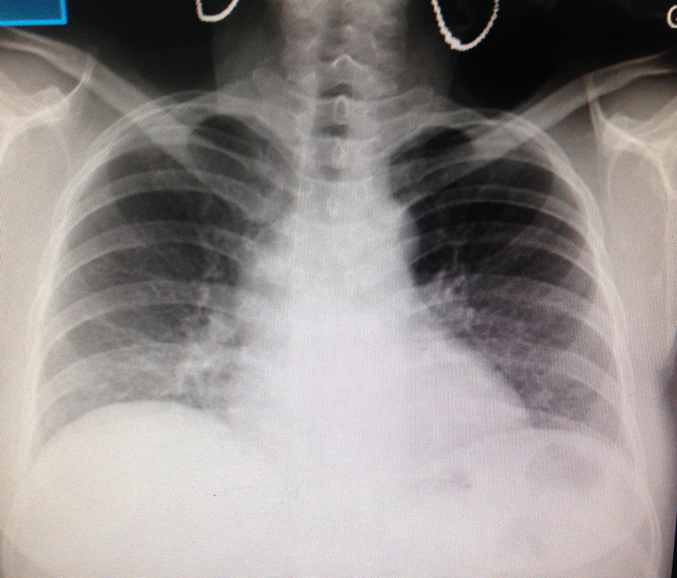
Clinical examination ruled out  surgical needs , chest  X-ray is normal.


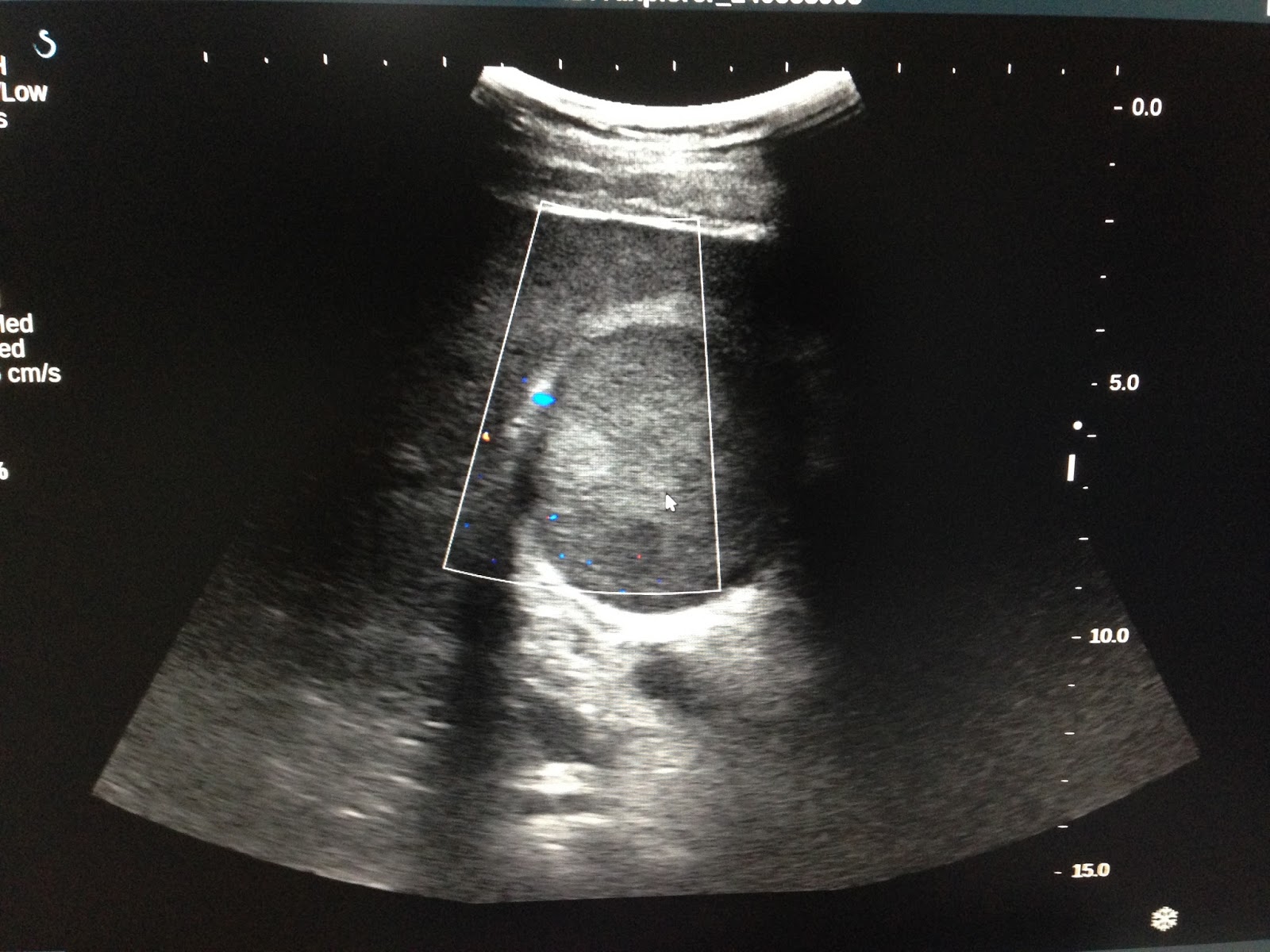
  • Ultrasound of abdomen = liver, biliary system,  kidney are normal, huge amount of  ascites volume with cloudy fluid  (us1 , us2, us3 pelvis  us4 ovary).


CT scan of abdomen  =  No tumor detection [ ct1,frontal view, ct2, ct3  cross section].


Blood tests =  WBC  15k with 13,3 k neutro, CRP= 25.9,  amylase, CEA , CA 125  are  normal level.  But Widal test is  positive th;1/320
Ascites punction= yellow  clear,   analysis =  ADA= 19.5 ng/mL,  CA125 :396 UI/mL, CEA: 0,8UI,  albumine =3.9 mg/mL


After 2 weeks treated with antibiotics;   response is good,   no fever  but ascites  is  distention after  aspiration  many times.
Summary=   Fever with typhoid  but ascites  still  persistent after one month.

Discussion :   Fever and   Widal test positive  is  Typhoid fever, treated response with antibiotics, but ascites  is still  progressing, so that  is not feature of Typhoid fever (one  expert of infectious disease says). Ascites analysis  can rule out  pancreatitis,  tuberculosis and  cirrhosis.  With CT , ultrasound  don't  detect any tumor intra abdomen. Then the short  way for diagnosis is  laparoendoscopy  for biopsy.


Laparoendoscopy  detected  multiple white spots intra parietal peritoneum,  most common in diaphragma area  (see foto)  suspected peritoneal carcinomatosis.


Microscopic result is malignant mesothelioma.


Reference:
Xem ca 164