Total Pageviews

Tuesday 4 July 2017

CASE 441: LIKE CHERRY SKIN TUMOR, Dr PHAN THANH HẢI, MEDIC MEDICAL CENTER, HCMC, VIETNAM


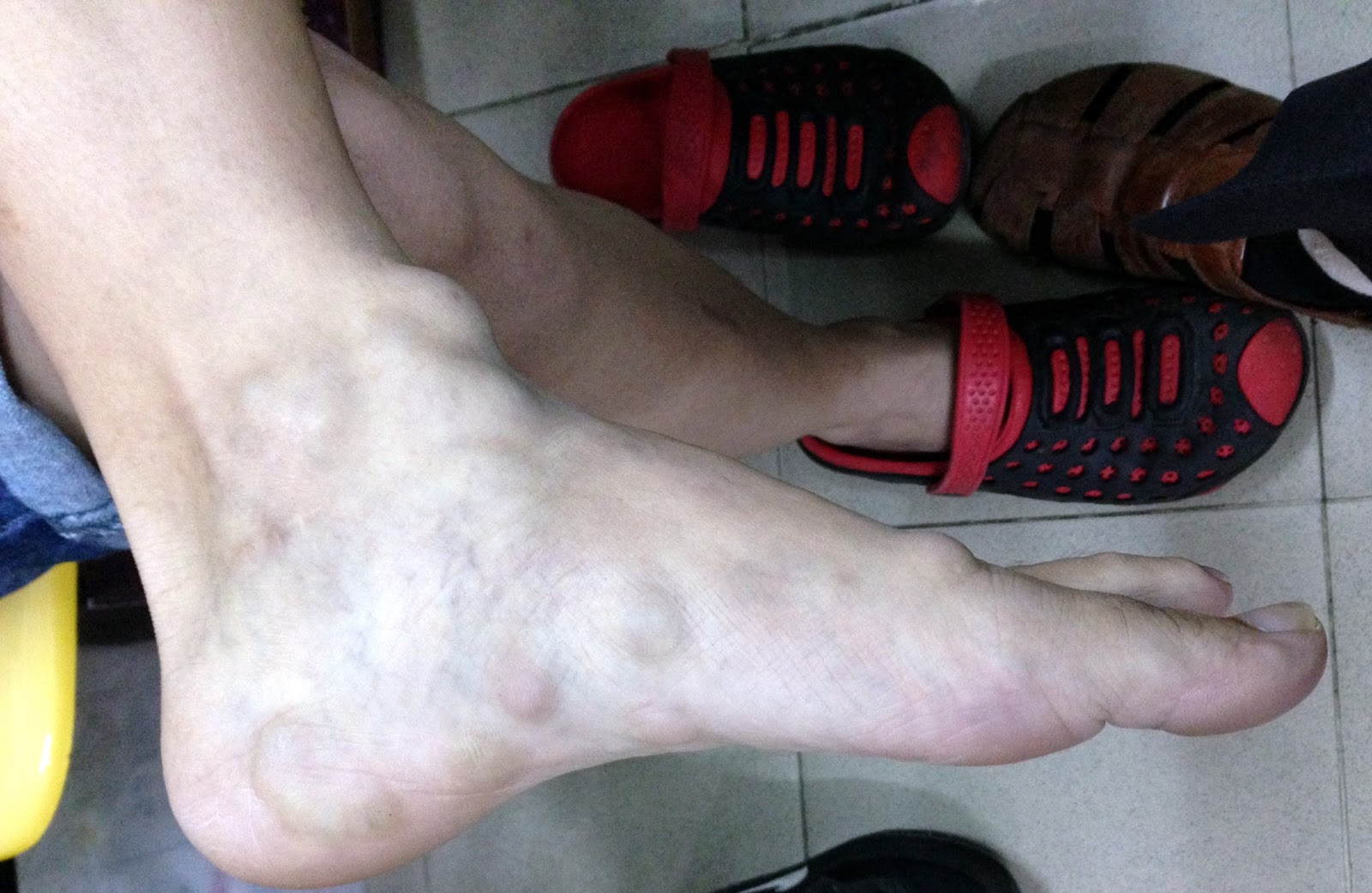
Lady 17 yo  with tumor  on her left shoulder   which was  detected for  2 weeks,  size of 3 cm, red and soft,  no pain ( see photo 1,2).


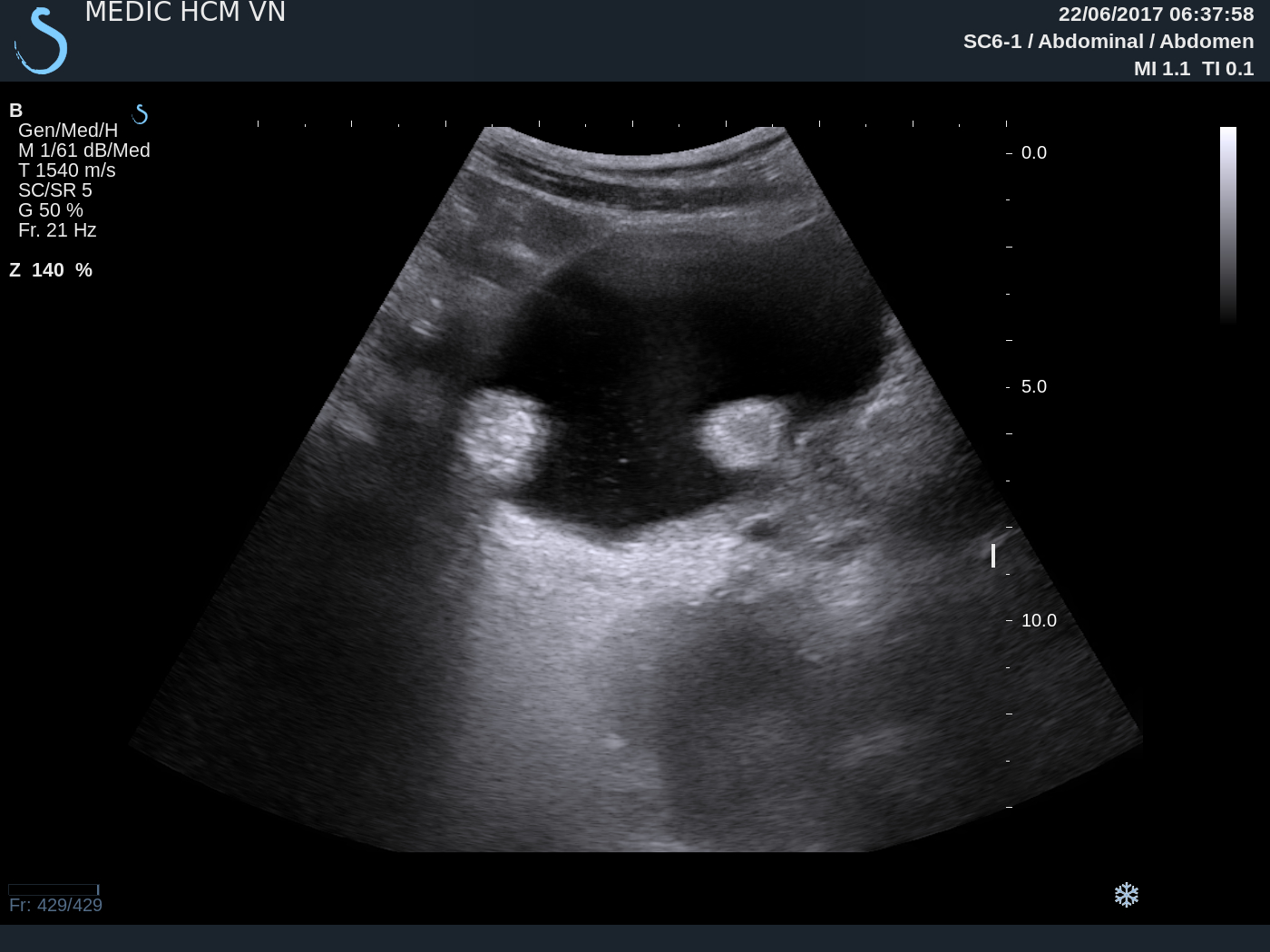
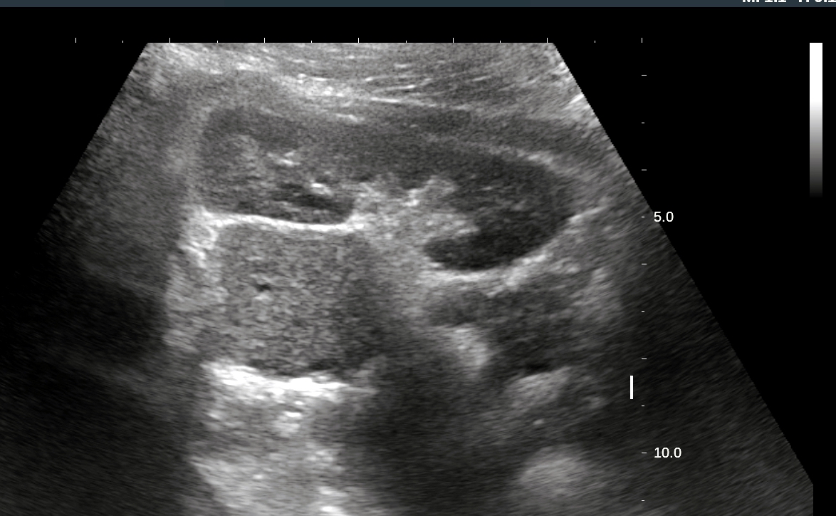
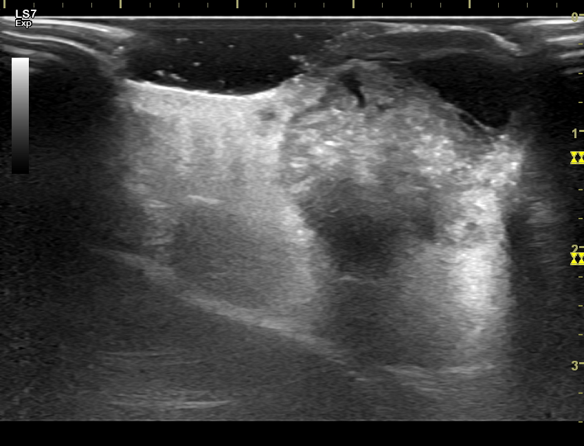
On ultrasound examination,  this tumor  came from  skin layer, inhomogenous with solid part in the root and calcification, while upper part is fluid (US 1, 
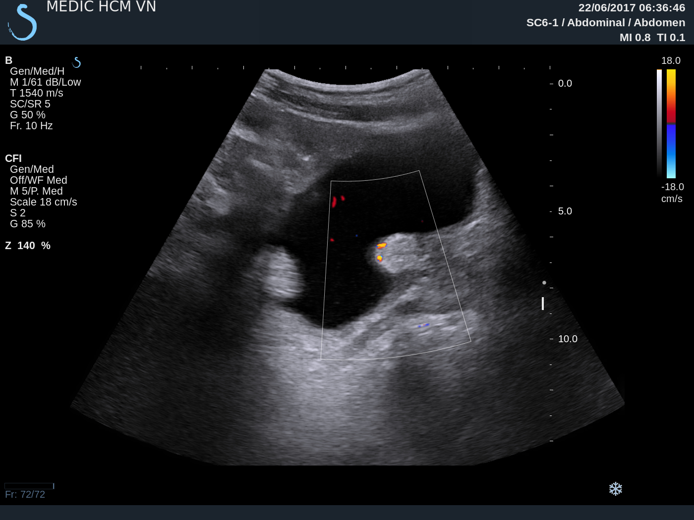
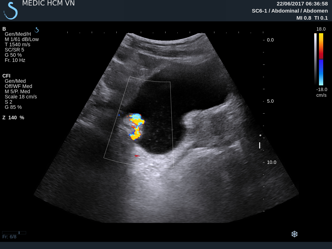
US 2). On CDI, tumor  has hyper vascular pattern like an octopus.


OPERATION  REMOVED THIS TUMOR.


MICROSCOPIC RESULT IS MALHERBE'S disease  
[ PILOMATRICOMA ).

Sunday 2 July 2017

CASE 440: MULTIPLE TUMORS of the LEG, Dr PHAN THANH HẢI, MEDIC MEDICAL CENTER, HCMC, VIETNAM


Man 20 yo with  history of 10 years ago having many small subcutaneous  tumors on left  foot ,  size of 2 cm, no pain. And now he detected another nodule near his left knee (see photo1, photo 2).


 It  is soft in palpation, no pain,  compressible and reexpansion after releasing it.
Ultrasound examination of  this tumor showed  tumor belonging to sapheneous vein while  deep vein is normal.


MRI reported that tumor of superficial vein of left foot.


Biopsy of this small  tumor. Microscopic and histoimmuno staining is angioleiomyoma.


REFERENCE:

Thursday 29 June 2017

CASE 439: URINARY BLADDER TUMOR, Dr PHAN THANH HẢI, MEDIC MEDICAL CENTER, HCMC,VIETNAM


Man 69 yo with  hematuria. Ultrasound  detected 2 tumors in urinary bladder (UB), one in fundus of UB, another,  in prostate  ( US 1,  US 2 ), US 3=  color Doppler with twinkling artirfact due to calcification, US 4 = cystic tumor in left kidney.


MSCT with CE of  urinary tract confirmed left kidney cystic tumor and urinary bladder tumor.


By via cystocopy biopsy result is transitional cell carcinoma of UB.


Tuesday 20 June 2017

CASE 438: RENAL HILUM TUMOR, Dr PHAN THANH HẢI, Dr NGUYỄN VĂN AN, MEDIC MEDICAL CENTER, HCMC, VIETNAM


Female patient 21 yo detected high blood pressure of 17/10 cmHg.
Ultrasound of abdomen detected  one mass at  upper pole of left kidney with  size of 3.5 cm. This mass  covers  left border of aorta, left renal hilum and adrenal fossa (US 1, US 2, US 3).


MSCT with CE= CT 1, CT 2,  CT 3, CT 4 (3D vascular)=    this tumor  covers the hilar kidney, very high CE enhancement. Radiologist says  adrenal tumor.


Blood test=catecholamine blood and  24 hrs urine analysis detected nothing abnormal
Metanephrine  blood =102 unit  (n= 90),  in urine = low 42 unit.
Operation  by laparotomy=

 Picture 1= this tumor  covers  the  left renal hilum.


Picture 2= Nephrectomy with tumor specimen.


MICROSCOPIC REPORT WITH  IMMUNO HISTO CHEMYSTRY  STAINNING IS  NON FUNCTIONAL PHEOCHROMOCYTOMA .


REF CASE REPORT


Sunday 11 June 2017

CASE 437: LIVER TUMOR, Dr PHAN THANH HẢI, Dr VƯƠNG THỪA ĐỨC, MEDIC MEDICAL CENTER, HCMC, VIETNAM


Female patient 16 yo with epigatric pain.

Blood test=  HP positive.
Ultrasound  detected  left liver tumor.

US 1 =  longitudinal scanning of  left lobe of liver:   solid tumor , size  of 10 cm with central necrosis.

US 2 =  subcostal scanning :   tumor covers left liver lobe.

US 3  = color Doppler :  vascular supplying  of  this tumor.


US 4 =  elastoscanning of this tumor = 10,5-15 kPa.  


MSCT with  CE: CT 1, CT 2 =  artery and vein phases, CT 3 =  frontal view of this tumor from left liver.


Blood tests=  no infested HBV, HCV, Wako tests 3 negative.


PRE OPERATIVE  RADIOLOGIST AND SURGEON  SAY THAT  LIVER FOCAL NODULAR HYPERPLASIA (FNH).

OPERATION WAS DONE FOR RESECTION OF THE TUMOR (SEE  MACRO).


Macroscopic is  FNH.


Liver tumor FNH or HCC


Reference: Macro FNH and  flow chart of identification FNH-HCA-HCC.