Total Pageviews

Monday 17 October 2016

CASE 399: HCC GOING TO HEART, Dr PHAN THANH HẢI- Dr NGUYỄN TUẤN VŨ, MEDIC MEDICAL CENTER, HCMC, VIETNAM


MAN 66 yo with CHEST PAIN and DYSPNEA. EMERGENCY ECHOCARDIOGRAPHY DETECTED  INTRA CARDIAC MASS, LOOKED LIKE THROMBUS.
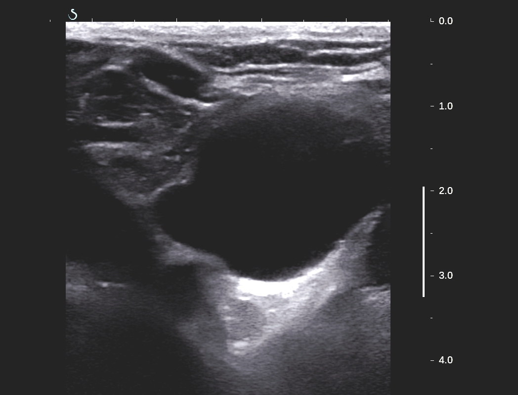
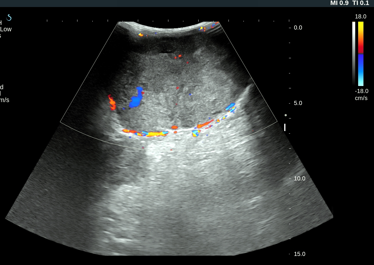
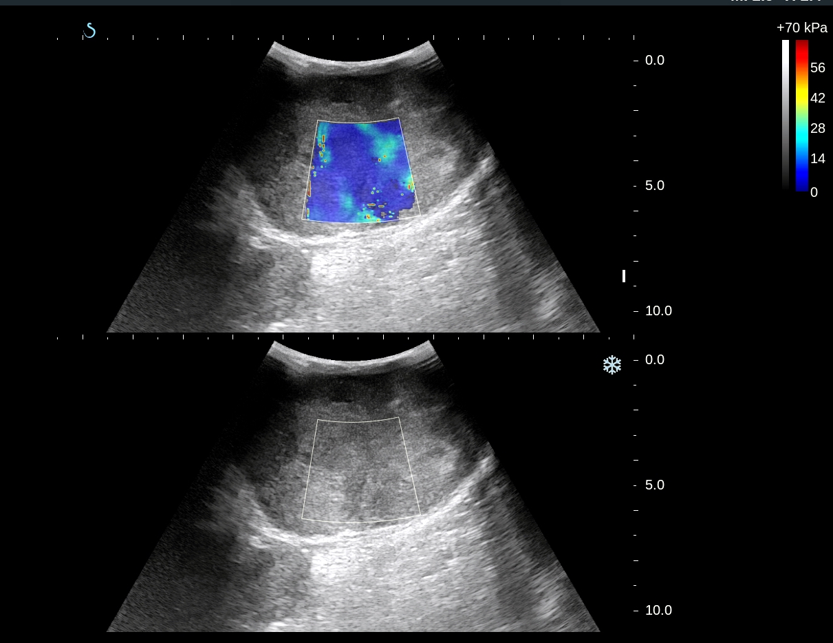
ULTRASOUND SCAN of LIVER DETECTED a LIVER MASS SIZE of 4cm and DILATED 2cm in DIAMETER and OBSTRUCTED by A HYPOECHOIC MASS ( US 1, US 2).
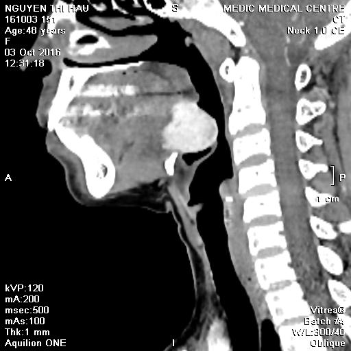
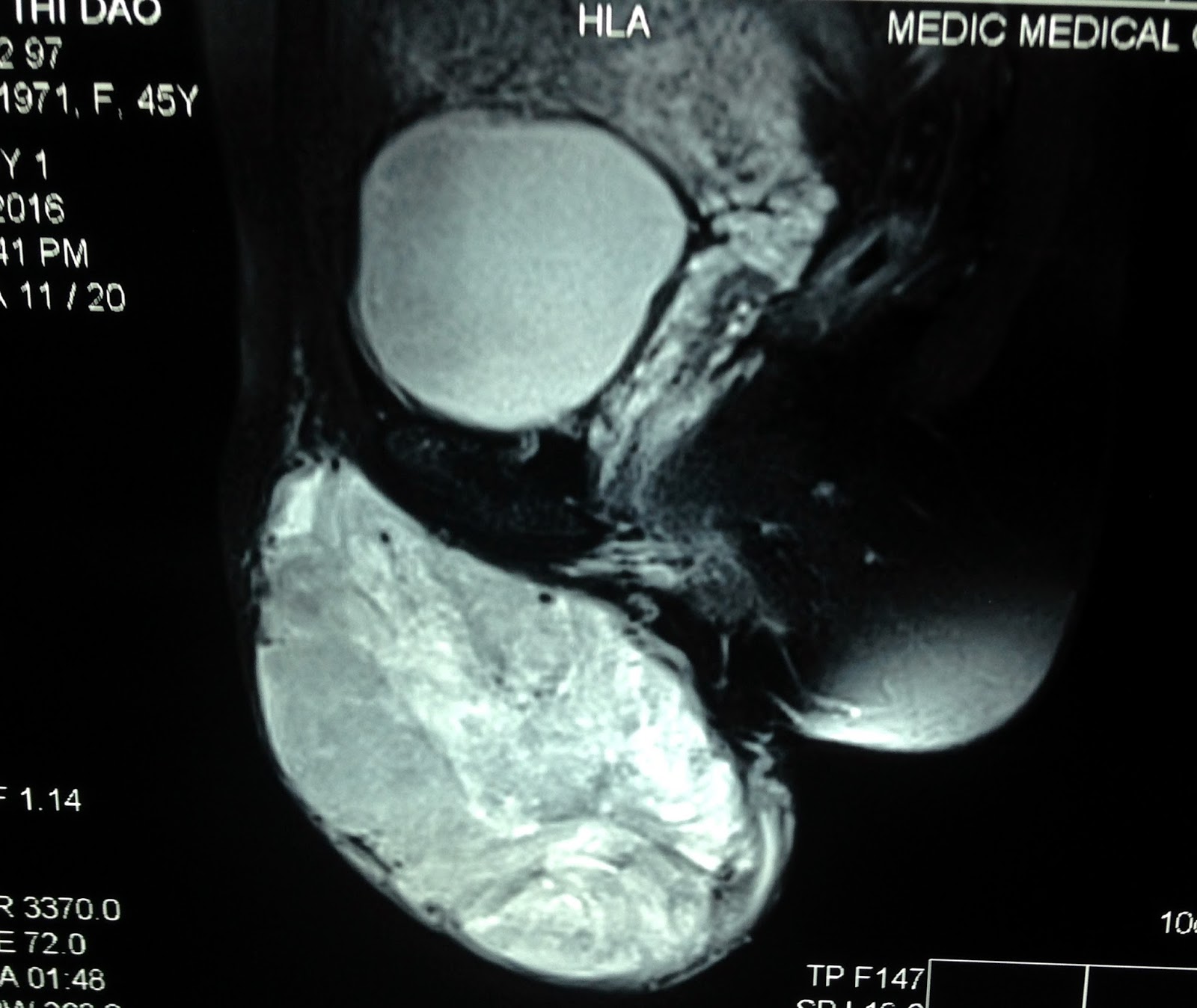
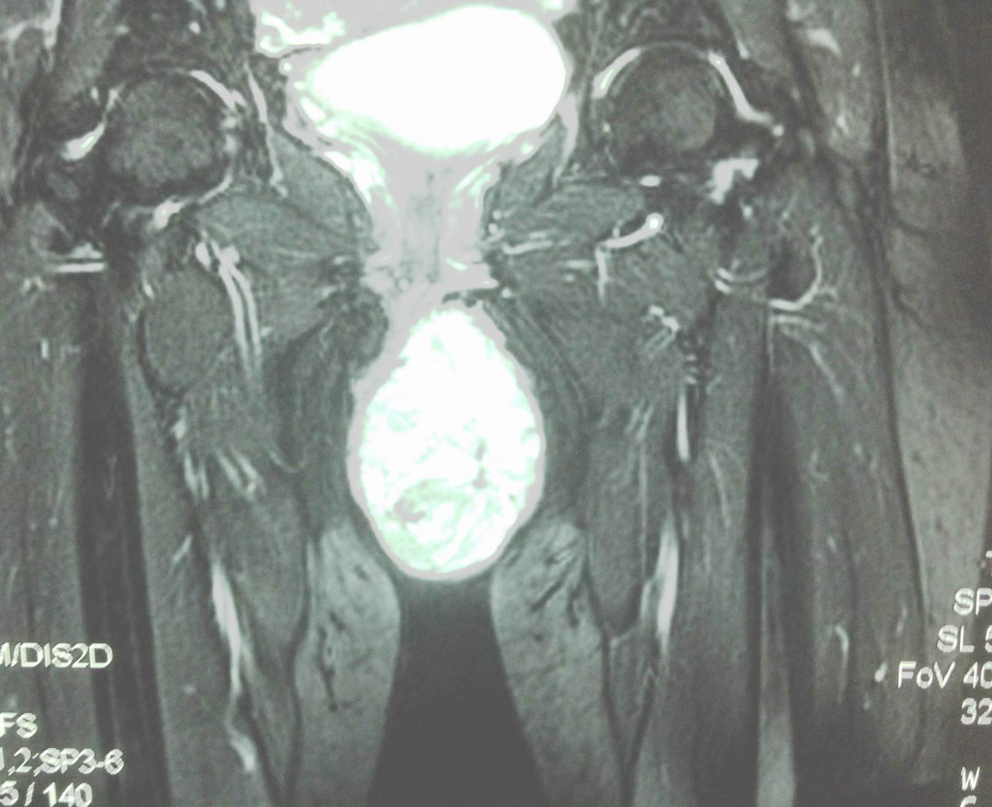


MSCT with CE DETECTED LIVER MASS and TUMOR INVASION TO HEPATIC VEIN TO IVC and GOING to RIGHT ATRIUM and FILLING DEFECTED at PULMONARY ARTERY (CT1, CT2).
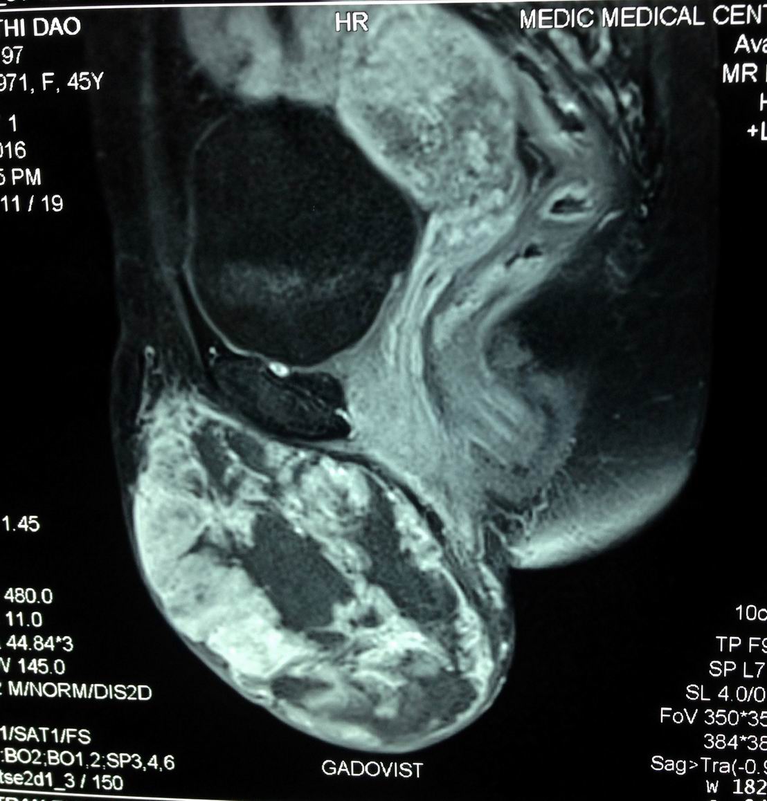


CT3 (section) SHOWED LIVER MASS, RIGHT ATRIUM MASS and INTRA LEFT VENTRICULAR MASS.


BLOOD TEST = HCV POSITIVE; WAKO TEST = TRIPLE POSITIVE.

Tuesday 4 October 2016

CASE 398: ECTOPIC THYROID GLAND, Dr PHAN THANH HẢI, Dr TRẦN THỊ BẢO CHÂU, Dr NGUYỄN THIỆN CHÂU


Woman 48 yo, PARA 2002, in general check-up, sonologist  detected no thyroid gland at normal location (US 1).


Blood tests reported  TSH= 11.47 microIU/mL (n= 0.3-5 microIU/mL),  Free T4= 0.985 ng/dL ( n= 0.7-1.85 ng/dL).
Ultrasound  again..detected  one mass  at  basal tongue,  size of 3x4 cm, hypoechoic,  hypovascular  (US 3, US 4).


MSCT with CE  detected  intralingual  mass which is  very quick  enhanced of  CE, size of  4 cm (CT1, CT 2, CT3).


CTA  detected  no thyroid arteries both  superior or inferior branches from the CCA and  ECA ( CT 4)


Conclusion=  Ectopic thyroid gland  intra basal lingual type.

Reference
reference case 319 medic link case-319-lingual-thyroid  

Saturday 1 October 2016

CASE 397: NERVE TUMOR, Dr PHAN THANH HẢI, Dr NGUYỄN NGHIỆP VĂN, MEDIC MEDICAL CENTER, HCMC, VIETNAM.


Boy 17yo with pain at anterior brachial region both 2 sides. Clinical palpation revealed many small subcutaneous nodules, size as a pepper nut, painful at palpation ; and swelling neck (see chest XRays).


Ultrasound scanning of the antebrachial areas detected many small ellypsoid nodules along the arterial way looked like the nerve ( US 1, US 2: longitudinal scan and cross-section).


US 3, US 4 scan at the carotid detected hypoechoic mass along the carotid sites  which belong to vagus nerves 2 sites.


US 5, US 6 scan at left carotid= big size vagus nerve, hypoechoic #1.5 cm in diameter at cross section.


MRI of the neck and thorax= MRI 1, MRI 2: hypertrophic nodular vagus nervi go to  mediastinum.


MRI 3: lateral view of the neck; MRI 4 = cross section of vagus nerve at the neck in relation with carotid artery.


MRI 5: hypertrophic intercostal nerve at thorax.

Biopsy of  the subcutaneous nodule at forearm  reported  neurofibroma in microscopic result.


Monday 26 September 2016

CASE 396: PARATHYROID CYST, Dr PHAN THANH HẢI, Dr NẠI HƯƠNG THOANG, Dr VŨ TU THÂN


Woman 43 yo with  sorethroat  and cough, sputum bleeding.
Chest X-Rays for  screening: no  chest lesion ( see foto chest X-Rays).


Ultrasound of  the neck: normal thyroid  but detected a cyst at lower pole of thyroid gland, size of 5-6 cm, monocystic  prolonged to retrosternum.
US 1,US 2 ( CDI),   US 3    pretrachea longitudinal scanning.


MSCT CE of  the neck: CT 1=frontal viewing,  well bordered cyst, CT 2: sagittal view..
C T 3. Cross-section= retrosternum tumor.


Ultrasound guide punction  of this cyst removing 10ml clear fluid.
What do you need to study in this fluid?

Fluid analysis report: high PTH  78.69pg/ml  and in the blood PTH is 47pg/mL.

The final diagnosis is  non functional PARATHYROID  CYST.

  
Reference


Monday 19 September 2016

CASE 395: MAJOR LABIA TUMOR, Dr PHAN THANH HẢI, MEDIC MEDICAL CENTER, HCMC, VIETNAM


WOMAN 45 YO, SAYS  THIS VULVA MASS  IN  SLOW GROWING 6 MONTHS AGO, WITHOUT  PAIN ( FOTO).


ULTRASOUND OF THIS TUMOR REVEALED SOLID, FIRM,   SIZE OF 10CM.
 US 1:WITH  B MODE,    US 2 WITH  CDI : HYPOVASCULAR SOLID TUMOR /  US 3=ELASTOULTRASOUND OF THIS TUMOR IS HARD STRUCTURE.


MRI 1 SCAN = SAGITTAL SECTION OF THIS TUMOR
MRI 2 WITH  GADO  CE= IN LATE PHASE, LOOK LIKE  A CYSTIC DEGENERATION.
 MRI 3 = FRONTAL  SECTION OF THIS MASS  FROM LEFT MAJOR LABIA,  PEDUNCULATED  LOOK  LIKE SCROTUM IN MALE PATIENT.


Core  biopsy reported  microscopic with  immmunochemistry staining is  glomus tumor.


Operation remove this tumor (see macro)


.
Reference:

https://www.scitechnol.com/glomus-tumor-in-vulva-with-uncertain-malignant-potential-1Mbz.pdf