Total Pageviews

Friday 22 January 2016

CASE 360: RIGHT KIDNEY TUMOR, Dr PHAN THANH HẢI, MEDIC MEDICAL CENTER, HCMC, VIETNAM.


Man 38yo 2 years ago  intermittent  hematuria, today  acute right renal colicky pain. 
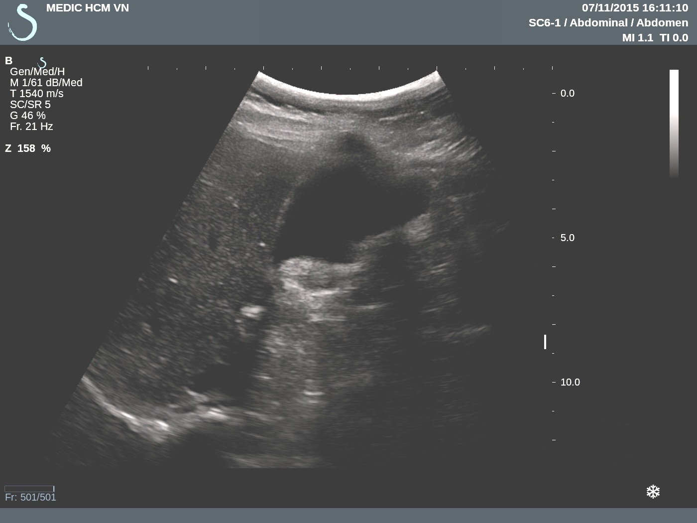
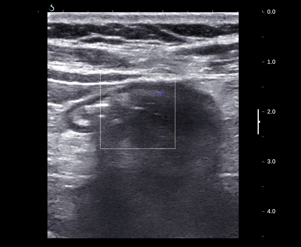
Ultrasound in emergency detected big right kidney and fluid collection arround  kidney.


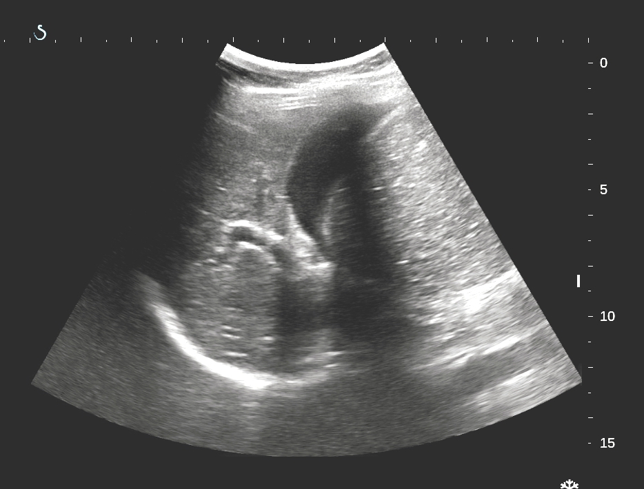
Pelvic kidney  has a collected  hyperechoic mass which made  dilated ureter.
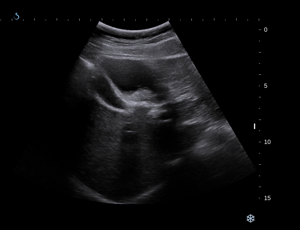
CDI ultrasound detected  no Doppler signal in  vascular renal cortex ( US 2)

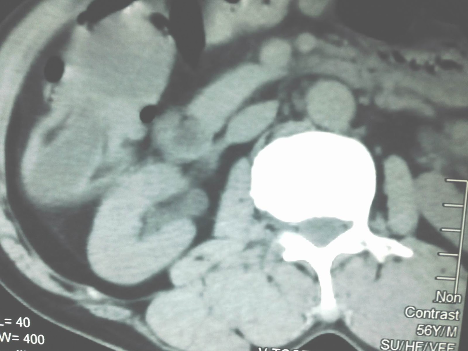
MSCT with CE=CT1: frontal view=  right kidney  too big without contrast  supplying.


CT 2: frontal view, pelvis of right kidney  is covered by enhanced contrast mass  just to dilated ureter. 


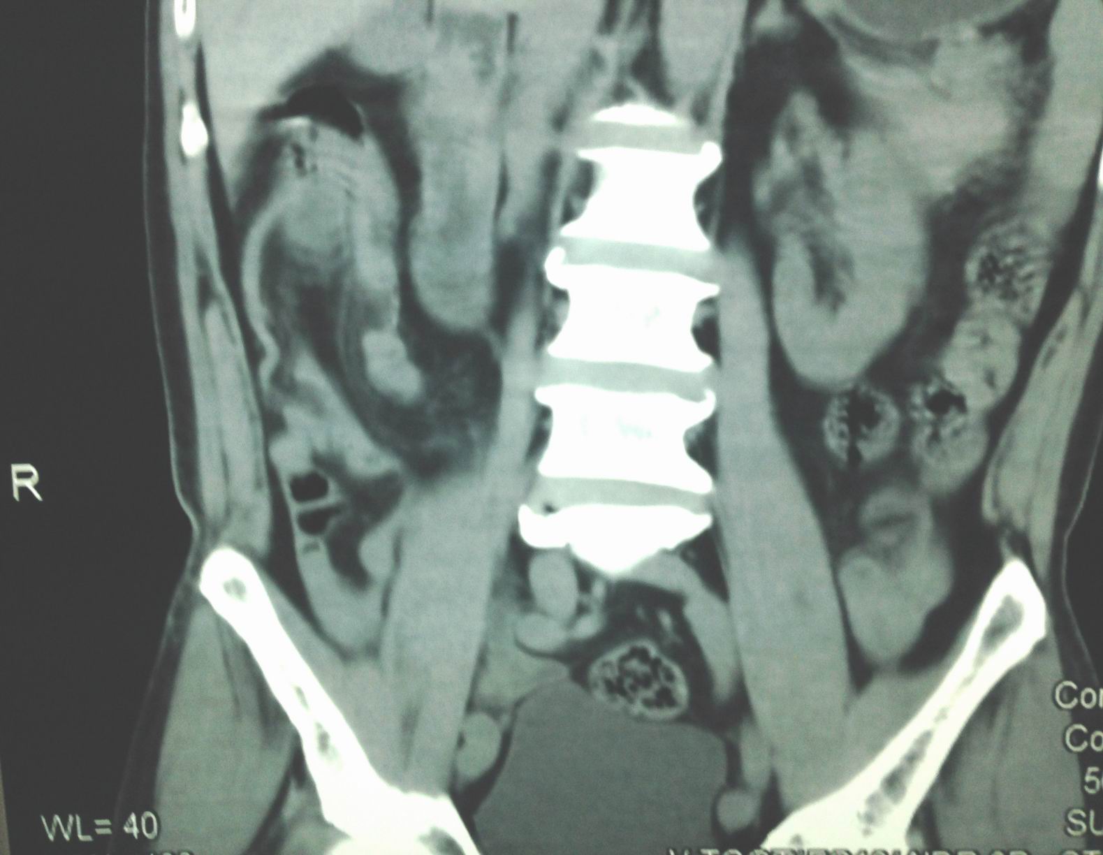
CT3, CT 4: cross- sectional view: pelvis and ureter detected  intralumen  one enhanced  contrast structure  liked  a tumor.


CT 6: 3D vascular view= no vascular supplying to right kidney.


Report  by radiologist  is  bleeding intra  right urinary system with  ureter obstruction  by  tumor, suspected  TCC.(TRANSITIONAL CELL CARCINOMA)
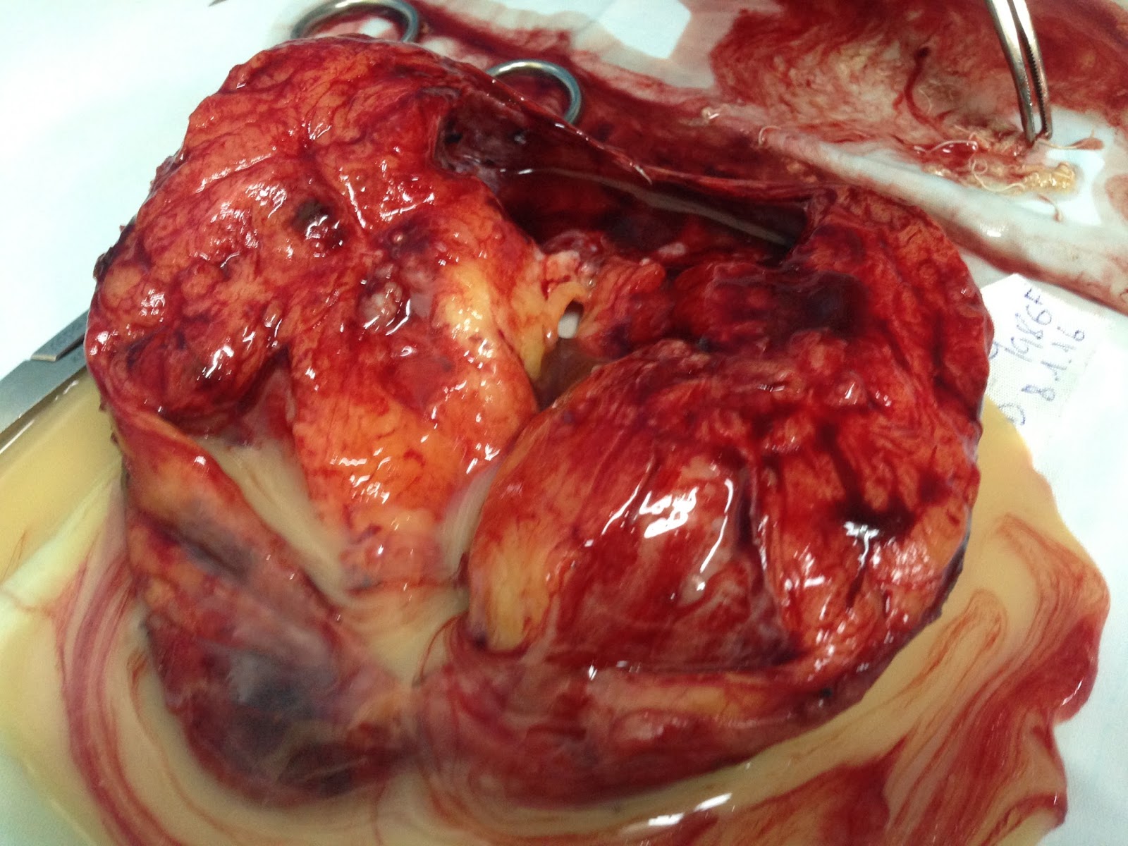
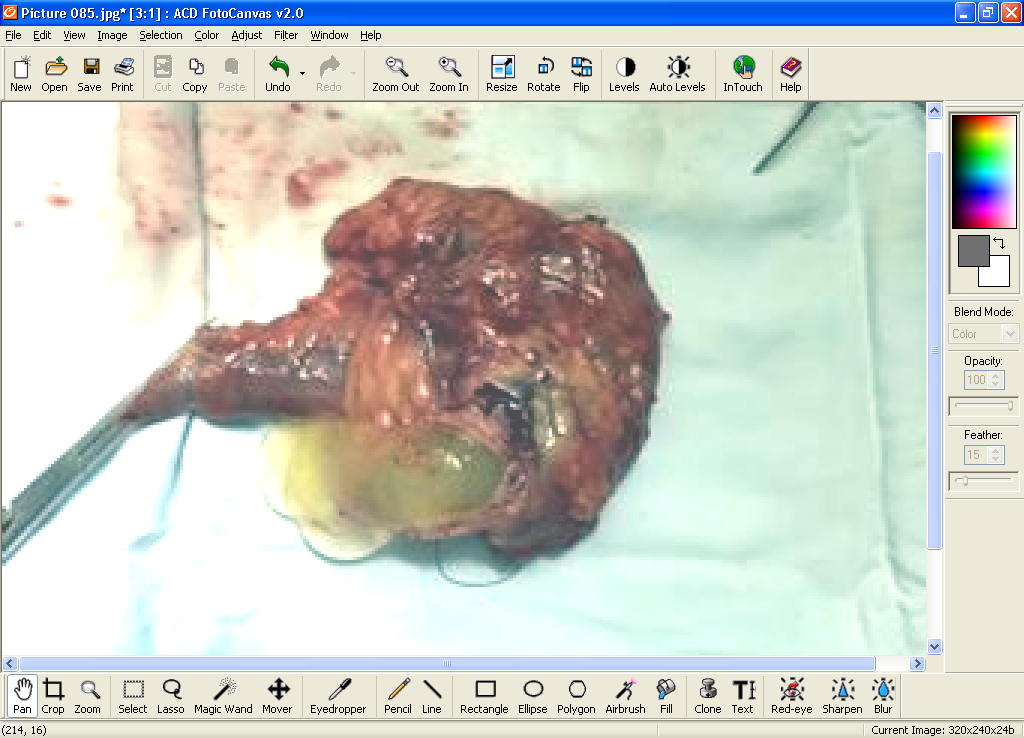
Emergency operation  of right nephrectomy and  ureterectomy.
Macroscopic specimen showed  tumor in obstruction of distal ureter.


Microscopic report TCC  ( transitional cell carcinoma hight grade malignancy.


Thursday 14 January 2016

CASE 359: RIGHT HIP PAIN, Dr PHAN THANH HẢI, MEDIC MEDICAL CENTER, HCMC, VIETNAM.


Women 72 yo, pain at right hip  in walking for 2 months , no trauma, no fever.
Ultrasound of right hip joint ( us 1 scan,  us2, us 3  cross- section).


Plain XRay in AP view  for comparison of right to left hip joint ( XRay image)  no abnormal detected.


CT scanning  ( CT 1 : cross section of  head of  femoral  bone deformation  at right side, CT 2: frontal view,  CT3  3D view).


MRI  of hip joint  in comparison of  right to left  femoral head bone.


Final diagnosis is AVN ( avascular necrosis of femoral head)

Saturday 9 January 2016

CASE 358: LESSER OMENTUM TUMOR, Dr PHAN THANH HAI , Dr VAN UYEN, MEDIC MEDICAL CENTER, HCMC, VIETNAM


Women 30yo, general check- up .
Ultrasound detected a tumor on border of liver near gallblader which deplaces left gastric curvature and is from retroperitoneal space. Its structure are solid and cystic parts,  size arround 10cm ( see ultrasound  us1.. cystic part tumor  in border  liver; us 2..near gallblader;  us 3..long scan  left lobe liver and tumor.). Sonologist cannot  diagnose this tumor  from lesser omentum.


MSCT with CE of this tumor  is mixed structure, cystic, fatty, and calcification [ CT1..section, CT 2  frontal section , CT3  sagital ). Suggession from radiologist  is teratoma tumor or  lipoma necrosis.


MRI   with gado ( MRI 1..struture is more fat tissue., MRI 2..with  fat suppression ,  MRI  3 frontal view).  Radiologist  says  teratoma of retroperitoneum, in lesser omentum area.


Blood test  of all  cancer markers are normal.
Laparo-operation= 


picture 1( retrogastric tumor well bordered)
picture   2macro
picture  macro 3, opened specimen,   solid and cystic tumorand  fluid inside  like milk)

Microscopic report of this tumor is teratoma maturation.


Friday 1 January 2016

CASE 357: PELVIC MASS, Dr PHAN THANH HẢI, MEDIC MEDICAL CENTER, HCMC, VIETNAM.

Female  15 yo, pain at pelvis, no mentrial cycle.
Ultrasound of  pelvis   one  sonologist  suggested  ovarian tumor.

Review of  ultrasound:

US1:   cystic structure in pelvis, long 20cm, morphology like a  hourglass, upper portion  near  bifurcation of abdominal aorta.


US2:  cross-section..of the mass :  fluid and debris inside.


US3.  Cross- section,  the wall of the upper part  is thiskening.


US4 with linear  probe:  wall is   thickening  in comparison to  the lower part.


Ultrasound   report suggested  a  hematometriocolpos.
MRI of pelvis detected  this mass with  old blood  inside.


Diagnosis is  imperforated hymen  and  hematometriocolpos.
ObGyn doctor resolved it by  incision of hymen to drainage  this old blood.


Monday 28 December 2015

CASE 356 : COLO-COLIC INTUSSUSCEPTION, Dr PHAN THANH HẢI, Dr VÕ THỊ THANH THẢO, MEDIC MEDICAL CENTER, HCMC, VIETNAM


Man 56 yo, acute  colic pain at  right  upper quadrant of  abdomen,  crisis and  vomitting 3 days ago.
Ultrasound  scanning of liver

US1: Ultrasound detects big mass  near gallblader liked a bowel loop dilated.


US 2: Right colon dilated  with  multiple layers  which is oignon sign.


US3. Coecum moved  up near liver  connected with one cystic mass.


US 4  Cystic mass  with  multiple  rings  [oignon sign],  typical of  mucineous cyst  of appendix.


CT scan abdomen detected  right colon  moving up  with  coecum  intussusception (CT1 frontal  section;, CT2, sagital section; CT3, frontal section).


Emergency  operation with diagnosis  colo-colic intussusception  by  appendicular mucocele.
See specimen of operation by right colectomy.


Microscopic report  is appendicular mucocele. 
Reference:

Saturday 26 December 2015

CASE 355: PAROTID GLANDS TUBERCULOSIS, Dr LÊ ĐÌNH VĨNH PHÚC, MEDIC MEDICAL CENTER, HO CHI MINH CITY, VIETNAM


A female 19 yo patient, student, swelling and pain in the parotid glands about a week, not fever.


Ultrasonography showed multiple structures within the parotid glands on 2 sides, hypoechoic, well-defined, measuring approximately 5 - 12 mm, with the umbilical node. She was diagnosed inflammation of the parotid glands and antibiotics for ten days  (cephalosporin 3 and fluoroquinolon).


But parotid glands swelling continuosly, ultrasound images  with more nodules in the parotid glands,and antibiotics for ten days again. In next follow-up visit parotid glands biopsy was done, and result showed chronic salivary gland inflammation.
Patient was sent to hospitalization Ho Chi Minh city in dentomaxillofacial center for 2 weeks of antibiotics as Sjogren syndrome. Parotid glands still  swollen and had discharge line to detect skin.  And she returned to MEDIC for parotid gland ultrasound.

Ultrasound image showed multiple hypoechoic structures with fluid inside, well-defined, proliferative vascular supplying, created road detect skin.

MSCT with CE showed parotid gland hypertrophy, having multiple lesions with fluid density in the central area.


Parotid gland biopsy showed salivary gland with Langhans great cells.


Parotid gland fluid examination showed high ADA and PCR/ TB (+).