FOR PICTURES PLS CONNECT TO 3G / DOWNLOAD THE LINK
Man 52 yo, fever
unknown origine for 3 months, blood
tests: nothing abnormal detected.
MSCT
scan of full body detected a small
nodule on right lung, size of
1 cm with some pericarena lymph nodes
enhanced with CE and one subcutaneous
mass of 3 cm in the back of left neck (CT
lung images).

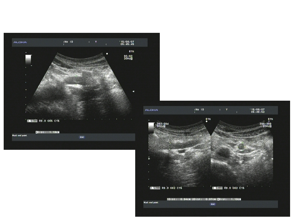
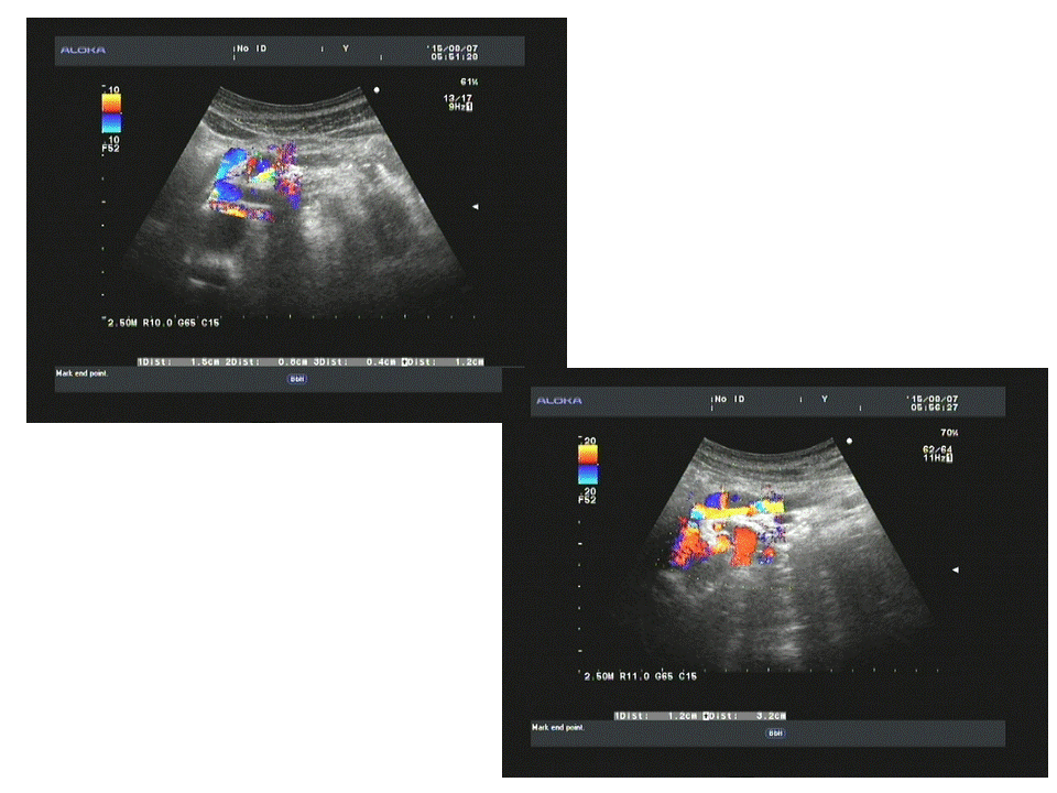
Ultrasound
of this mass revealed round
border, very hypoechoic, nonvascular
filling intramass, no posterior enhancement, no sister mass together ( see 3 US
images and video clip).


Biopsy was done for this
mass and microscopy result was
adenocarcinoma metastasis from the lung.

Discussion: Clinical onset is fever unknown origine, CT lung detected small spicular nodule , pericarena nodes and the patient himself detected one subcutaneous mass at posterior of his left neck; biopsy of this mass made diagnosis of metastasis from lung cancer which is small cell lung cancer.
Conclusion = Small lung cancer metastasis to skin and
paraneoplasic fever.
Reference: Case in NEJM.