Total Pageviews

Tuesday 16 June 2015

CASE 317:CT FIRST U/S SECOND IN CAUTION OF THYROID FUNCTION, Dr PHAN THANH HẢI, MEDIC MEDICAL CENTER, HCMC, VIETNAM

FOR PICTURES PLS CONNECT TO 3G/ DOWNLOAD THE LINK


MAN 29YO  TRAUMA  AT THE HEAD AND NECK BY TRAFFIC ACCIDENT, NOW IN  PAIN  AND TACHYCARDIA.

CT  
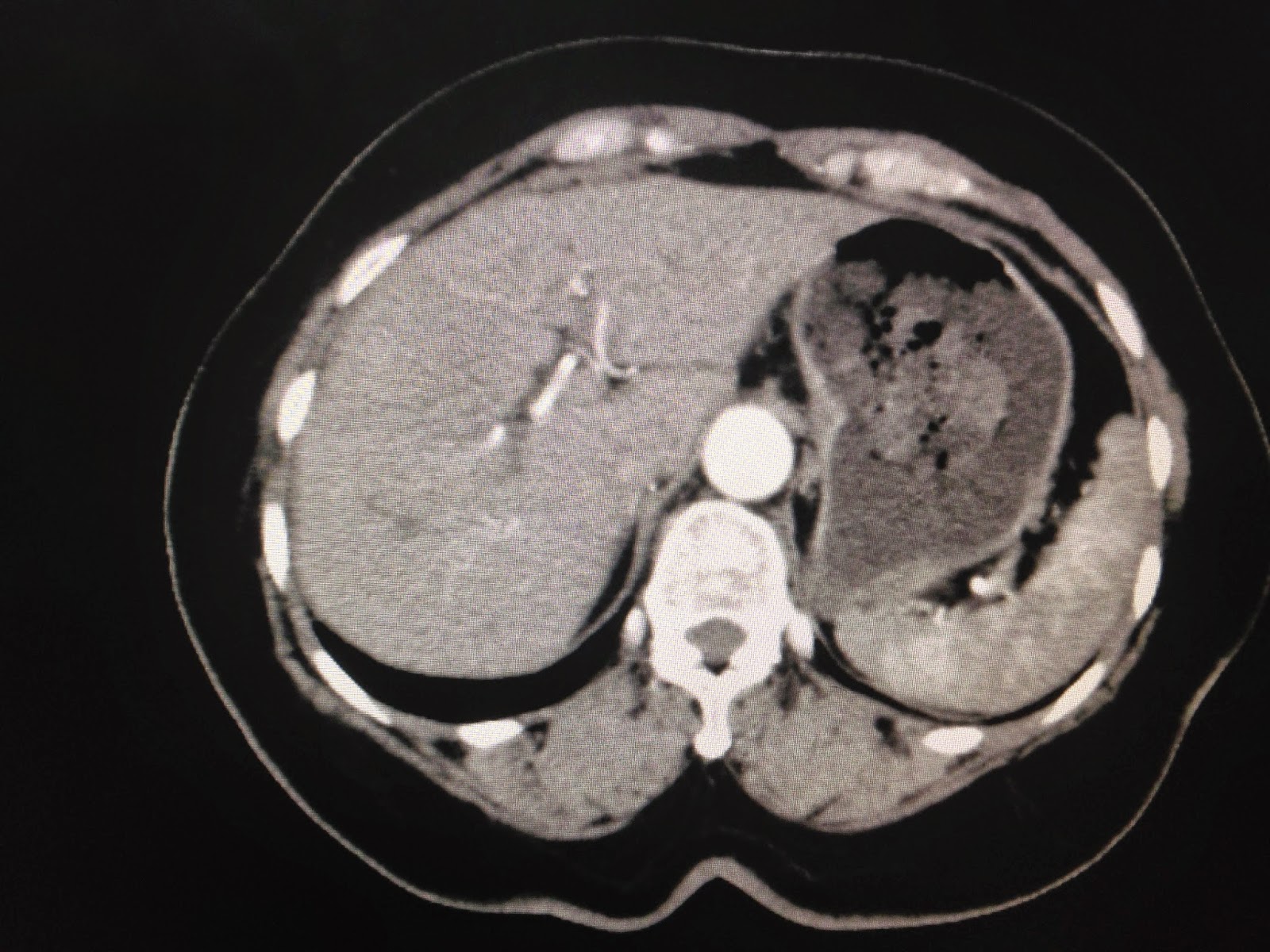
NON -CE WAS FIRST LINE, RADIOLOGIST  REPORTED    BIG AND  DARK THYROID GLAND , CT UNIT ARROUND   85 UI HOUSFIELD ( SEE  FOTO AND  CT1,CT2 ARE  ABNORMAL).


 ULTRASOUND  OF  THYROID  ALSO    REPORTED LARGE VOLUME WITH  HYPOECHOIC AND   VERY  HIGH  SIGNAL DOPPLER . SPECTRAL DOPPLER  OF  SUPERIOR THYROID ARTERY  WAS VERY  FAST PULSATILE, TYPICAL HYPERTHYROIDISM  AS  GRAVE  DISEASE (BASEDOW).


BLOOD TESTS  CONFIRMED  A  HYPERTHYROIDISM  WITH TSH  LOW  VALUE  AND VERY  HIGH T3 AND T4.


SUMMARY: CT  NON CE  IS  POTENTIAL IN CAUTION  THYROID FUNCTION.


Sunday 31 May 2015

CASE 316: RETROPERITONEAL CALCIFACATED TUMOR, Dr PHAN THANH HẢI, MEDIC MEDICAL CENTER, HCMC, VIETNAM

 FOR PICTURES PLS CONNECT TO 3 G / DOWNLOAD THE LINK


WOMAN 45 YO, PAIN  AT  RIGHT  SUBCOSTAL REGION. PLAIN XRAY FILM  KUB DETECTED  CALCIFICATION  PUNCTIFORM   AT LIVER BORDER AND  RIGHT KIDNEY ( SEE XRAY PLAIN FILM).


ULTRASOUND  OF ABDOMEN  FIND OUT  THIS MASS  IN RETROPERITONEUM,  SIZE  OF 7X5 CM , WHICH PULL THE RIGHT KIDNEY DOWN. ITS STRUCTURE  IS  CYSTIC,  MULTILOCULATED WITH  FINE WALL  AND  CALCIFICATED. THE CONTENT OF THIS CYST IS  MIXED ECHOGENIC STRUCTURE ( SEE  4 US IMAGES ).


MSCT OF ABDOMEN  WITH CE=  THIS TUMOR IS  MULTICYSTIC  WITH  CALCIFICATION OF  THE WALL ( SEE 3  CT IMAGES).


BLOOD TESTS  AND  MARKERS = AFP,  BETA HCG ARE NORMAL


Preoperative  diagnosis  in  BD HOSPITAL IS retroperitoneal teratoma.
Operation  removed  this mass   hard  and clear bordered, specimen  is  white  structure  like  fibroma.


Microscopic result is fibroma in retroperitoneum.
REFERENCE:


Monday 25 May 2015

CASE 315: SPONTANEOUS PORTO-SYSTEMIC SHUNT, Dr PHAN THANH HAI, Dr NGUYEN THI ANH HONG, Dr LE THONG NHAT, Dr TRAN LAM

FOR PICTURES PLS CONNECT TO 3G / DOWNLOAD THE LINK

case 315

Woman  63 yo,  5 years ago  had been treated  diabetes  not  control blood  sugar. Patient  has  some  subcoma  crisii that had been treated in many  hospitals, now she  is in somnolence, easy in sleeping  after eating but cannot sleep at night.
MRI of  the brain, radiologist  detected  hyperintense  T1 at basal ganglion area, susgested  hepato-encepalopathy (image MRI).


Checking  the liver by blood tests, liver function is still good,  but ultrasound of  liver  detected  porta-systemic shunting  very high flow ( see US image 1 dilatation cystic  intrahepatic, US 2, color Doppler:  porta-systemic shunting and US video).


MSCT angio of  liver with  3 phases: arterial phase shows that not abnormal ; venous  phase : dilated  the  porta-systemic  anastomosis like snake. In 3D CT reconstruction  confirmed a  high flow porta-systemic shunting).


Bood test reports  this time  no ceton, high NH3 = 88.89 micromol/mL (normal  18-72).
Conclusion: It is  a high flow  spontaneous  porta-systemic  shunting appeared as hepatic encephalopathy in clinical examination.

REFERENCE:

Saturday 23 May 2015

CASE 314:CALCIFIED LIPOMA of CALCANEUS TENDON and SOLEUS MUSCLE - - Dr PHAN THANH HẢI- Dr LÊ THANH LIÊM- Dr Nguyễn Đức Duy Linh; Dr Nguyễn Ngọc Xuân Giang – BÌNH AN GENERAL HOSPITAL_KIÊN GIANG_VIỆT NAM.

FOR PICTURES PLS CONNECT TO 3 G / DOWNLOAD THE LINK

case-314-lipoma-calcification-of-calcaneus-tendon-and-soleus-muscle-

A 30 year-old woman with history of mass within the right heel region 2 year ago, no pain  and normal walking.
ULTRASOUND
Ultrasound found an echogenic mass in the calcaneus tendon and soleus muscle, hypovascular.
The surface of the mass was close to the Achilles tendon, well-defined with the capsule. Deep surface was very difficult to survey. Intra the mass there was an hyperechoic structure with strong shadowing, like bone. Ultrasound did not determine that there was continuity between the mass with calcaneus tendon or not.
Sonologist suggested a diagnosis of chondro-osteoma and organized synovial cysts of the ankle. Dx: Lipoma calcification.

CT SCAN
MSCT detected a lesion in the soleus muscle, density of fatty tissue, 10x4cm in size, around of achilles tendon, clear capsule, rounded border, with a very high-density nodule inside, looked like the bone. The lesion was not associated  with the calcaneus bone and the leg bone. Radiologist suggested a diagnosis of calcified lipoma.


OPERATION
Surgery was conducted in next day. Macroscopic image looks like the lipoma with calcified nodule inside.


Microscopic result is benign lipoma.


REFERENCE:

CASE 313: FISH BONE APPEARING in NECK, Dr PHAN THANH SƠN, Dr NGUYỄN ĐỨC DUY LINH, Dr NGUYỄN NGỌC XUÂN GIANG, BÌNH AN HOSPITAL, KIÊN GIANG

FOR PICTURES PLS CONNECT TO 3G / DOWNLOAD THE LINK

case-313-fish-bone-appearing-in-NECK

Male patient 79 yo had foreign body on right neck (see pictures).
HISTORY
4 days before of hospitalization, during dinner with stingrays, in sneezing, a 79 yo patient took out and in some fish parts, and then feeling caught something in his throat but still eating.
Morning after he went to hospital as felt felt like a head of bone at his right neck.


ULTRASOUND

 Ultrasound at right neck revealed a fish bone in soft tissue, echogenic line in subcutaneous edema around the fish bone, but no vascular.

ENT ENDOSCOPY


 ENT endoscopy detected edema of right piriformis recess due to fish bone perforating outside from the throat.

CT SCAN
CT–Scan confirmed the fish bone in the right neck, not injured the right vessels of the neck.


The fish bone was removed out of the right neck, with 2.8 cm long of size.


Fish food is very interesting, but sometimes lead to dangerous situation like the case, specially for old patient. 

REFERENCE;


Monday 18 May 2015

CASE 312: A Case of PENA-SHOKEIR PHENOTYPE: FETAL BODY HYPEREXTENSION, Dr PHAN THANH HẢI, Dr JASMINE THANH XUÂN, Dr ĐẶNG VĨNH PHÚC, Dr TÔ MAI XUÂN HỒNG

Antenatal ultrasonography findings and magnetic resonance imaging in a case of Pena–Shokeir phenotype Xuan-Hong Tomai, Thanh-Xuan Jasmine, Thanh-Hai Phan

case-312-fetus-hyperextension

Female 33yo with cutaneous allergy from age of 20, now in 3rd pregnancy. 2 first girls are normal and no abnormal family history. She has been treated  allergy while was  in 12 weeks of gestation [medrol, cefixin, antihistamine) for one month and changing to Prednisone pommade.


She was recorded the 3rd pregnancy with 2 OB ultrasound examinations at 12 weeks and 18 weeks: Fetus with normal attitude, heart rate 158b/min, nuchal translucency NT=1.1mm,  normal fetal vertebral column.


But  the 3rd OB ultrasound examination, in 23th week, detected a  micrognathia, abnormal  curved attitude of vertebral column, hyperextension,  which appeared in days of follow-up. 2 small lungs in small thorax, a few movements of 2 upper and lower limbs and fingers in extension time by time in uterus.  But other viscera  of fetus were normal.


MRI detected loss of septum pellucidum.


The parturient was sent to HV hospital for a prenatal care, and  an amniotic fluid analysis was done . FISH test showed no abnormal chromosome of 13th, 18th, 21th , X and Y,  and karyotype result in 4 weeks next. In one week follow-up in hospital, with many sonologists, fetal  attitude no change which made thorax  more compressed, small stomach and polyhydramnios due to fetus cannot swallow.
Because of  nothing change for one week,  the poor pregnancy had to be end.


DISCUSSION
Fetus in 23 weeks with malformation= abnormal hyperextention of vertebral column and four limbs and micrognathia, no septum pellucidum.  FISH test showed normal  13, 18, 21, X , Y chromosomes. (Double test and Triple test  not yet performed).
In birth, micrognathia, fingers in gripping, curved vertebral column looked like abnormal 18th chromosome.


Few in literature of abnormal curved vertebral column of fetus, there was 2 papers but the vertebral column changes normal at birth without malformation, and babies remain well.