Total Pageviews

Wednesday 11 March 2015

CASE 301: SKIN METASTASIS, Dr PHAN THANH HẢI, MEDIC MEDICAL CENTER, HCMC, VIETNAM

XIN DOWNLOAD THEO LINK ĐỂ THẤY HÌNH
FOR PICTURES  PLS DOWNLOAD LINK BELOW.

MAN 83 YO HAD BEEN  TREATED  LUNG CANCER WITH  CHEMOTHERAPY  AND RADIATION  FOR 2 YEARS.
ONE  MONTH  AGO  HE DETECTED  SMALL SKIN TUMOR  AT LEFT TEMPORAL SCALP  RAPID  GROWING AND BLEEDING ( SEE PICTURE).


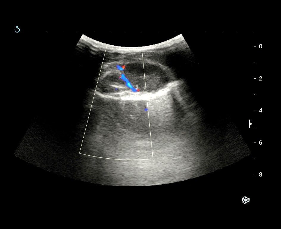
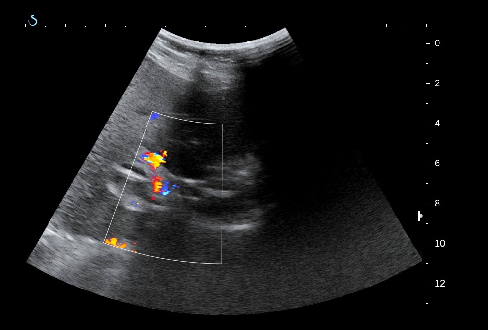
ULTRASOUND  SCAN   REPORTED  THIS TUMOR BEING  FROM THE  SCALP NO  INVADE  TO  BONE ( SEE  2 US  PICTURES)  AND   LIVER  METASTASIS ( US PICTURE 3)


MSCT  CONFIRMED THE  SCALP TUMOR   NOT ERODING THE BONE, MANY  METASTATIC  LESIONS FROM THE LUNG TUMOR  TO  BOTH SIDE OF LUNG, LIVER, ADRENAL GLAND.


BIOPSY OF THIS TUMOR  CONFIRMED THAT  METASTASIS FROM LUNG CANCER,  SQUAMOUS CELL CARCINOMA..


SUMMARY:   LUNG CANCER  SOMETIMES  METASTASES TO SKIN OF SCALP AND NOT TO BONE.

REFERENCE:


Friday 6 March 2015

CASE 300: MULTIPLE BONE TUMORS, Dr PHAN THANH HẢI, Dr HỒ CHÍ TRUNG, Dr LÊ THÔNG LƯU, MEDIC MEDICAL CENTER, HCMC, VIETNAM

XIN DOWLOAD THEO LINK ĐỂ THẤY HÌNH

case-300-multiple-bone-tumor

MAN 46 YO PAIN AT LEFT LEG  AT PALPATION. X-RAYS  DETECTED TIBIAL BONE EROSION.


ULTRASOUND  SCAN THIS MASS SHOWED CYSTIC FORMATION.( SEE US  PICTURES)


CHEST XRAY  FOR ROUTINE CHECK UP  PRE OP  DETECTED  ONE MASS AT  RIGHT THORACIC WALL..( CHEST XRAY).


ULTRASOUND SCAN THIS MASS WAS  SOLID MASS,  HYPOVASCULAR WHICH WAS FROM THE  RIB. (SEE US3..WITH 3.5 MHz,,US 4 WITH 12 MHz, US 5 CDI,  US 6  ELASTOSCAN).


MSCT of  THORAX: THIS  MASS WAS  FROM  THE RIB   AND  ANOTHER  MASS AT  RIGHT  LUNG SUSPECTED  CANCER.


BLLOD TEST REPORT=   CYFRA -21  HIGH.


CORE  BIOPSY  OF THORAX WALL MASS = METASTASIS TUMOR HISTO TYPE  ADENOCARCINOMA    SUSPECTED  FROM THE LUNG CANCER.


Tuesday 24 February 2015

CASE 299: PORTAL VEIN FOREIGN BODY, Dr PHAN THANH HẢI, MEDIC, MEDICAL CENTER, HCMC, VIETNAM

XIN DOWNLOAD THEO LINK ĐỂ THẤY HÌNH

case-299-portal-vein-foreign-body


Woman 65 yo, epigatric pain for one week, cannot eat  and  no fever.
Ultrasound of abdomen  in decubitus position  detected  vena porta thrombosis and some  white lines intra portal vein which  came from the wall of gastric  antrum (see 4  ultrasound  pictures  in ventral view).


For clear viewing of  portal  vein  we  scanned  the liver  by sitting position and dorsal view.


Portal vein  was in distension, no flow  due to  thrombosis, and  in crossed section of portal vein we detected a white foreign body.( 2  pictures  with  sitting position scan ).

MSCT with CE  for  evaluation portal vein found out  the  foreign body which  length of 5 cm  intra left  branch of portal vein and one another end was intra gastric antrum wall.
The foreign body was  covered by thrombosis intra  left branch of portal vein (see 3 CT  images).


Blood tests  confirmed  infection  with  rising WBC and high CRP, no  abnormal coagulation test.


With  the  past history of ultrasound  scanning in ventral and dorsal views, MSCT and blood tests, the first choice  of diagnosis was intraportal vein foreign body, which was liked toothpick in penetration the gastric wall and  entering  liver  to left branch of portal vein, that caused  portal vein thrombosis.

What is your suggestion and planning of treatment for the female patient?

FEEDBACK=

An anouncement about case 299 of MEDIC  on Google web after the case was posted  for 30 minutes.


Operation this case  by  open laparotomy detected   one  bonefish with length of 5 cm which penetrated the duodenum  to left lobe of liver and entering the  vena porta  left branche.


Removing bonefish and sutured  duodenum.


Monday 16 February 2015

CASE 298: Carcinomatosis Ascites, Dr PHAN THANH HẢI, MEDIC MEDICAL CENTER, HCMC, VIETNAM.

Woman  64 yo, abdomen distension slowly for 3 months (photo).


Ultrasound  first  found out ascites  with slouge fluid  and  scattered fragments, normal liver, omentum thickening  like cake with  many hypoechoic nodules  and  no tumor in pelvis.


Chest X-Rays  was  normal.


MSCT of abdomen reported  a large amount of  ascites and  great omentum  thickening  with  many  nodules,  enhanced with  CE  and no  ovary  tumor.


Punction of  yellowish  ascites  that cytology was  negative and  ADA negative. Blood test  was  very high CA 125.


Laparoendoscopy cannot detect  primary tumor, but many white  nodules  covered  the  great omentum  but not in parietal peritoneum.
Biopsy the great omentum nodule. Microscopic report  was  undiffentiated adenocarcinoma, suspected  come from GI TRACT or  OVARIAN CARCINOMA.


Discussion

Acites with  large volume is easy  diagnosed  by clinical and ultrasound.
Ascites  fluid analysis rules out  some common diseases.

In this case, CA 125 was very  high in the blood test,  but  CT scanning  cannot detect ovarian tumor.
Laparoscopy for  diagnosis and  biopsy made sure  the case being  carcinomatosis.
This case  also had  gastro-colonoscopic result  and CEA negative.

The  most  suggestion  of diagnosis for  this case is  PPSC  ( PRIMARY PERITONEAL SEROUS CARCINOMA) .
Wait for  histo immunostaining  report.

REF  CASE of  PPSC.


Tuesday 10 February 2015

CASE 297: CHRONIC BOWEL OCCLUSION, Dr PHAN THANH HẢI, MEDIC MEDICAL CENTER, HCMC, VIETNAM

Man 47yo,   history of  3 months  with  peristaltism crisii and severe pain at  RLAQ more and more.
Ultrasound  first showed that  small bowel  dilating in  hypermotion and ascites.


MSCT  detected  enterolysis, stenosis at ilium  pre-cecum and suspected tuberculosis. 


Medical  antituberculosis treatment was in setting up, but  onset  total  occlusion was  acute  in emergency.
Operation in emergency detected  one portion of  ilium in hard stenosis. 


Resection that portion and  microscopic result  is adenocarcinoma infiltrating the bowel.