Total Pageviews

Tuesday 20 January 2015

CASE 294: SIGMOID COLON VOLVULUS, Dr PHAN THANH HẢI- Dr LÊ THỐNG NHẤT, MEDIC MEDICAL CENTER, HCMC, VIETNAM

Male patient 56 yo,  in emergency  going to MEDIC  by  acute  abdominal pain  and  distension. Clinical examination   this  patient cannot lay down ( see foto)


Ultrasound scanning of abdomen  first  detected  colon distension with air  and  hyperperistaltism (see 2 US images).


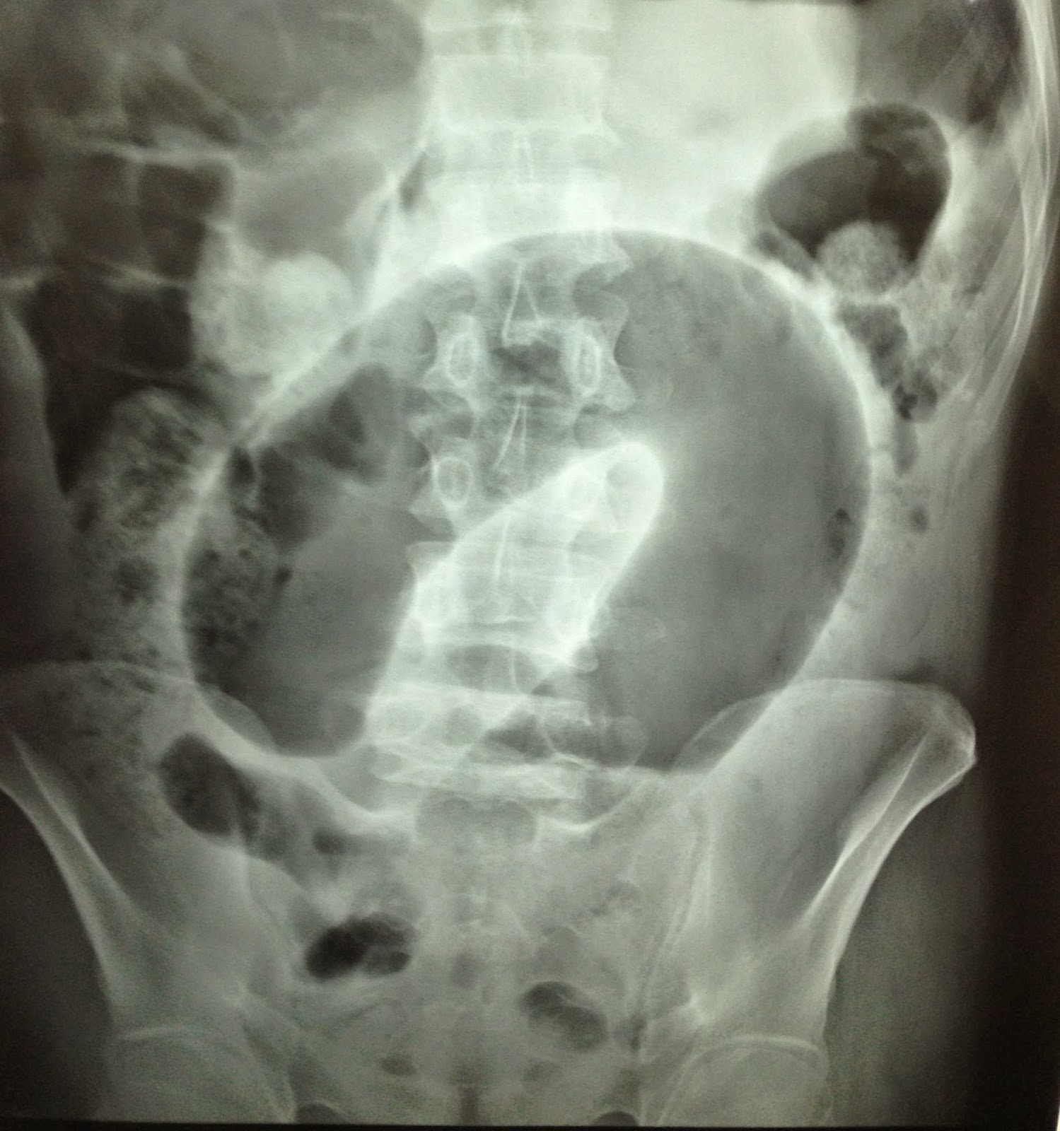
Next step,  a standing X-rays of abdomen was done  with  the  sign of C-loop, typical of  sigma colon  torsion (see   X-rays plain film).


MSCT of abdomen without CE presented  dilated colon  with air   (CT 1 double  black  ring  of  colon sigma distention,   CT 2: image  section of sigma colon  asymmetric,  CT 3:   image of coffee  bean, CT 4 : frontal section  with  mesocolon in torsion).


Radiologist  reported   volvulus of  colon sigma for the case.
Emergency  surgery detected  one part of  sigma colon  ischemic, resection  and colostomy with double canon technique.


Conclusion:  Emergency case  with ultrasound first choice for diagnosis, conventional x-rays  also can help patient  but  CT is the  best  information for this case.

Sunday 18 January 2015

CASE 293: KIDNEY ABSCESS, Dr PHAN THANH HẢI, MEDIC MEDICAL CENTER, HCMC, VIETNAM

Woman 58yo diabetes for 10 years, fever and epigastric pain radiating to left flanc.
Ultrasound of abdomen first detected one mass with air inside nearby left kidney which made bright line artifacts (see video clip), so that was thought to be a renal abscess.


MSCT with CE detected the abscess with air inside destroying lower pole of left kidney.


Blood test with WBC rising very high= 23,760 /mm3 with neutrophil 81.5%  and glucose urine=56mmol/L.


Ultrasound guided punction of renal abscess removed white pus. 


Bacteriology analysis result was  gram negative bacilli.
Treatment:antibiotic per IV  and surgical drainage of renal abscess and controlling  blood sugar by insulin.
Summary: Kidney abscess of diabetic patient was detected by ultrasound in recognizing gas inside abscess.

Sunday 11 January 2015

CASE 292: BREAST TUMOR of a YOUNG GIRL, Dr PHAN THANH HẢI- Dr JASMINE THANH XUÂN, Dr HỒ CHÍ TRUNG, Dr TRẦN THỊ HỒNG VÂN, Dr VÕ ĐĂNG HÙNG, Dr NGUYỄN SÀO TRUNG, Dr HỨA THỊ NGỌC HÀ, MEDIC MEDICAL CENTER, HCMC, VIETNAM

15yo patient from Dong thap province. For one month there was a small lump which was about one finger at right breast but it was getting bigger rapidly, and painless.


Right breast mass presented hypoechoic pattern, nearly took entirely breast volume, # 8cm x 10cm, hypervascular. Upon breast US the mass was inhomogenous, increasing diastolic phase and decreasing RI=0.37.


Mammo and MRI= Phyllod tumor or huge fibrosarcoma (BIRADS 3-4).


Core biopsy and immunohistostaining result: Malignant phyllod tumor or sarcoma of myxoid cellular stroma.


Radical mastectomy was done. Microscopic result was poor differentiated sarcoma.


Reference


Saturday 10 January 2015

CASE 291: ABDOMINAL PAIN POST CESAREAN, Dr PHAN THANH HẢI, MEDIC MEDICAL CENTER, HCMC, VIETNAM

Woman  34 yo, very painful some days during menstrual cycle for 2 years ago and after  cesarean operation  3 years before. Pain at  10cm  upper  pubis   near middle line and clinical palpation detected  one  mass of round shape and pain  at pressure.
Ultrasound  scan detected this mass ellypsoid, size 4cm x 3 cm intra right  rectus abdominus  muscle.
With  probe  3.5 MHz  this  mass was hypoechoic pattern looked like a cyst,  with  a small artery inside (see image 1, and video).


With linear probe 15MHz  this mass  had inhomogeneous structure, central  necrosis. Elastoscan this mass was  soft at central part (see  image 3 and 4).


FNAC was done for diagnosing the mass. Result from pathologist was hematoma with cells that were suspected endometrium cells.


What are your guidings for treatment for the patient?

Monday 5 January 2015

CASE 290: PARACERVICAL SPINE ABSCESS: Dr PHAN THANH HẢI, MEDIC MEDICAL CENTER, HCMC, VIETNAM

Woman 30 yo presented cervicalgia and swollen neck.

Chest Xrays: one mass at right of her neck.


Ultrasound detected thyroid gland normal( US1), but retrothyroidal spaces both 2 sides had hypoechoic masses, no vessels  intra mass which were suspected abscesses ( see  US 2:cross-section  at  lower  part of the neck, and US 3:longitudinal scanning of the neck).


MSCT of the neck (CT1 image showing the normal thyroid gland and 2 mass at lateral cervical spine looked like abscess). Frontal  view  and  sagital view detected  osteolytic lesions  at thoracic spine T1.


Ultrasound guided aspiration pus from abscess and detected BK positive in pus analysis.

It was a POTT' abscess of the neck, due to  T1 spine tuberculosis.

Tuesday 30 December 2014

CASE 289: GRAPE'S SIGN in LIVER, Dr PHAN THANH HẢI, MEDIC MEDICAL CENTER, HCMC, VIETNAM


Man 27yo, one  month  ago, suffered from pain at  liver  with  low grade  fever.
First visiting of MEDIC ultrasound, he was detected at  segment 7 of liver  multiple  spots,  hypoechoic, size of 1-2 cm  with  the round border,  no rising vessels on Doppler. Elastoscan of these lesions was soft  tissue  as abscess. 


 Blood tests presented  WBC rising and parasite tests were  normal.


One  week later, with  more pain at liver, CT scan with CE made image  like  grapefruit as an abscess.
Blood tests:  WBC rising  and  more eosinophil, CRP and IgE rising.


Discussion:
Based on clinical  signs  and  blood tests rising eosinophil ( 1.05 /n, <0.5 x10⁻⁹ ), CRP =  62.23 , IgE=670) and liver US  and  CT scanning with  multiple abscesses  like  grapefruit, we suggested parasite  abscesses  of liver, but  ELISA test not positive with any common parasite.
TREATMENT  IS  FOCUS  ANTI PARASITE LIVER ABSCESS.

After  2 days of ANTIPARASITE TREATMENT patient had  cough  and  pain at  right chest.
Chest Xrays  detected infiltration  lesion at  upper  pole  of right lung.
CT of lung  with  CE suspected  tuberculosis  with pleural effusion..


Puncture  aspiration of the pleural fluid that was clear yellow color.
Pleural fluid analysis : ADA was low value, NOT CORELATED WITH  TUBERCULOSIS.


Wait for  PCR result later.