Total Pageviews

Friday 20 June 2014

CASE 263: BUFFALO'S NECK, Dr PHAN THANH HẢI, MEDIC MEDICAL CENTER, HCMC, VIETNAM

Man 27 yo, dental  pain  on right mandibular  for one week, he detected  the right side of neck along  SCM muscle getting hot and swelling, pain and fever (see photo).


He was treated  with antibiotics and went to ultrasound scanning. Sonologist detected this neck mass beeing   like abscess by fluid collection on right and left neck (see 3 photo and video).


MSCT found out the right mass along SCM muscle and one other mass on left side nearby thyroid gland.


Puncture this mass  removed the pus but  direct examination  with gram stain no bacteria. 
Operation for drainage. 


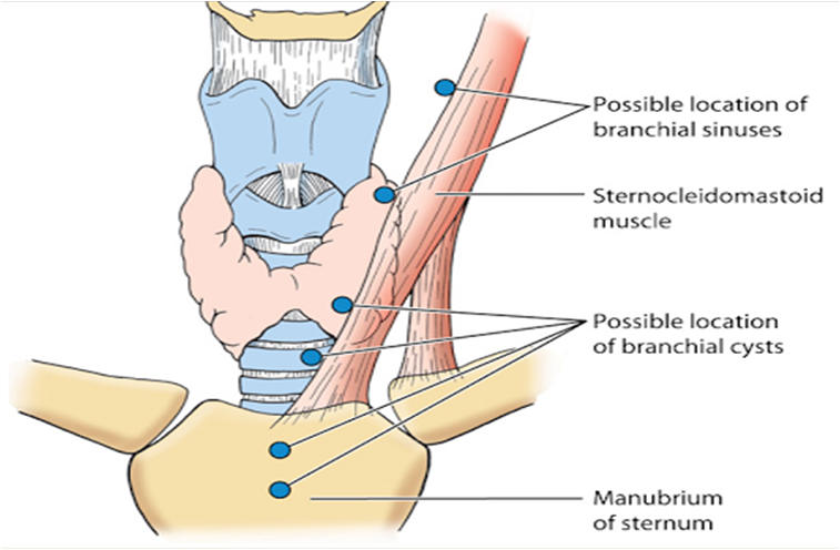
DISCUSSION: IT IS  A CYST of  INFECTION at LATERAL SITE  OF THE NECK. THE MOST COMMON IS  BRANCHIAL CYST.
CT  ALSO SUPPORTED THIS  DIAGNOSIS.

REF..ANATOMY OF  BRANCHIAL CYSTS.


Tuesday 17 June 2014

CASE 262: BIG ABDOMEN, Dr PHAN THANH HẢI, MEDIC MEDICAL CENTER, HCMC, VIETNAM

 Man 50 yo, one week ago, onset periumbilical pain and abdominal distension, no defecation nor fever.
Chest Xray, and  abdomen standing  plain film showed  the  water-air level in  intestine, suggesting  bowel obstruction.


Ultrasound  found out colon dilatation, filling water and moving circular around with hyperperistalsis (see video).


MSCT of  abdomen in  emergency detected dilated right colon and  small intestine, retroperitoneum edema  arround the pancreas and radiologist  suggested  that pancreatitis.


Blood test: WBC  rising 12k, amylasemia normal.
Operation  laparotomy detected  all  bowel in dilatation but  no  necrosis, no tumor obstruction. 
Many white spots like candle   intra peritoneum.
Retroperitoneal space edema. Surgeon said chronic pancreatitis.


Discussion of this case:  clinical findings were abdominal pain and distension for one week. XRay  and  ultrasound found out  bowel obstruction and CT  detected  pancreatitis, but  blood test amylasemia was 17 unit.
Surgeon decided operation by bowel obstruction.
Now  report  is  chronic pancreatitis, it is  a rare  case with normal amylasemia in acute  pancreatitis.

REFERENCE:  case report


Saturday 14 June 2014

CASE 261: HARD BREAST TUMOR like AMBARELLA FRUIT, Dr PHAN THANH HẢI, MEDIC MEDICAL CENTER, HCMC, VIETNAM

Woman 47 yo, she herself detected at the right breast one mass, slow growth, painless like one AMBARELLA fruit.
Ultrasound  first detected  this mass in size of 4 cm, at 10 am clock at  the right breast, with  many  white spots  as  calcification. Color Doppler finding also was a hypovascular mass.


Using elastoultrasound, the mass was  very hard,  scale  blue green color on elastogram, no detected  axillary  lymph nodes.


Mammography also detected  mass and microcalcification.


Core biopsy with ultrasound guided reports microscopic  INTRADUCTAL CARCINOMA, STAGING T2N0MX.


SUMMARY: BREAST CANCER IS  EASY DIAGNOSED BY ULTRASOUND   ELASTOGRAPHY of  THIS VERY HARD  TUMOR  LIKE  GREEN AMBARELLA FRUIT.


Wednesday 11 June 2014

CASE 260: ODDI TUMOR, Dr PHAN THANH HẢI, MEDIC MEDICAL CENTER, HCMC, VIETNAM

Man 40 yo  pain at  RUQ  with  dark urine.
Ultrasound  first   detected  big gallbladder  with  very think wall, no stone, CBD in dilatation just to   end  of CBD, no tumor of  pancreas (2 ultrasound images).


MSCT CE detected  dilated biliary system intra and extrahepatic ( 3 CT images).


Gastro-duodenal  endoscopy  found out a round tumor at Oddi  area (see photo),  biopsy was done.


Blood tests, CEA, CA-19-9 were normal.
What is  your suggestion for   diagnosis and your next step ? 

ERCP not successful.
Biopsy of the tumor but the microscopic result was negative.

Ooen operation for exploration, surgeon detected a hard massat the head of pancreas.
Whipple operation was performed  (specimen of tumor of Oddi).


Microscopic report was  adenocarcinoma ( patho images)


REFERENCE  from Meditoons


Friday 6 June 2014

CASE 259: PLEURAL EFFUSION, Dr PHAN THANH HẢI, MEDIC MEDICAL CENTER, HCMC, VIETNAM

Woman  23 yo  for one  month cough  and  dyspnea, no  fever.
Ultrasound of the thorax detected left  pleural  effusion with collapsed lung and  one mass covered  anterior mediastinum to external pericardium (see 3 ultrasound pictures of the left lung).


MSCT  with CE of   the  chest confirmed a  big  mediastinal  tumor   with  pleural effusion  which displaced  the heart to the right side (see 3 CT pictures).


Blood test and all  cancer markers  were normal. 


Transthoracic biopsy of this tumor with  ultrasound guided for immuno-histochemitry staining . Pathology report is  B CELL  LYMPHOMA.


This  patient is  going to treat with chemotherapy.

REFERENCE:   Mediastinal Tumor


Friday 30 May 2014

CASE 258: MALIGNANT G I S T RECURRENCE, Dr PHAN THANH HẢI, MEDIC MEDICAL CENTER, HCMC, VIETNAM

Man 54 yo,  2 years after operation for  acute obstruction of small  bowel by  tumor of  intestine, unknown microscopic  report.
Now he had  pain in the pelvis. (photo of  the skin scar operation).


Ultrasound  report   was  multiple  nodules  in  pelvis  looked like  grappe fruit
adherence  over  urinary bladder wall (2 ultrasound pictures).


MSCT CE of abdomen  detected   multiple  intramesenteric round tumors looked like  lymph nodes.


All blood test  and cancer markers were  normal.

Open laparotomy removed the mass which were  multiple  round tumors  adherent  to  great omentum and pedunculated (see  macro1, 2).


Microscopic report this tumor is  malignant  GIST recurrence.


Monday 26 May 2014

CASE 257: KNEE PAIN, Dr PHAN THANH HẢI, MEDIC MEDICAL CENTER, HCMC, VIETNAM

 Man 35 yo, 3 days ago, pain  at  right knee  cannot move, fever, no  history  of trauma. Clinical examination of right  knee:  hot and swelling at suprapatellar area (see photo).


Ultrasound first  scanned  at the right  knee, detecting  swelling of the  suprapatella recessus with homogeneous fluid  (2 ultrasound pictures).


MRI  of the  right knee  is  same   picture  report ( T1, T2).


Blood test  confirmed this infectious status  with  rising  WBC and  CRP.
For make sure  the  diagnosis: puncture of  the knee joint with  ultrasound guided..removing  the  yellowish  synovial fluid.
LAB analysis report  were  hight WBC and  negative gram stained bacteria  present in this fluid.


Diagnosis of  this case is acute  bacteria  infection of  the knee  joint, emergency  treated  with  antibotic and analgesic drugs.
DISCUSSION: in acute  case  ultrasound   guided  puncture of the  joint is fast action  for fast  diagnosis.

REFERENCE: