Total Pageviews

Thursday 15 May 2014

CASE 254: CASTLEMAN DISEASE in COLONIC MESENTERY, Dr JASMINE THANH XUÂN, Dr PHAN THANH HẢI, MEDIC MEDICAL CENTER, HCMC, VIETNAM

A 22 yo female patient with a  mass of right abdomen which was detected by ultrasound check-up and thought to be a mesenteric tumor or a lymph node in mesentery. It was well-bordered and vascular structure without any symptom.
MSCT confirmed the 14x17mm mesenteric tumor in right abdomen with CE enhancement.


Open surgery removed the mass from posterior space of right colonic mesentery.

Microscopic result is a Castleman disease in mesentery,  which is an uncommon lymphoproliferative disorder that may be localized to a single lymph node (unicentric) or occur systemically (multicentric).


It was a  second case at Medic Center.
The first case of Castleman disease  was posted in 2010.
CASTLEMAN DISEASE in RETROPERITONEAL SPACE at MEDIC CENTER


Saturday 10 May 2014

CASE 253: GOSSYPIBOMA (TEXTILOMA) POST CAESAREAN SECTION for a YEAR, Pham Hong Dong,M.D; Nguyen Duc Duy Linh,M.D; Phu Van Tuot,M.D; Nguyen Ngoc Xuan Giang,M.D., MEDIC Binh An Kien Giang Hospital


A 26 year-old female patient who had complained mild pain at her pubic region  presented lower abdominal pain a month prior. She  overwent a caesarean section a year ago for delivery her child.
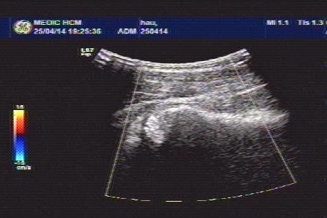
Ultrasound findings:A cystic mass (about 83x46 mm) containing distinct internal hyperechoic wavy, striped structures.    


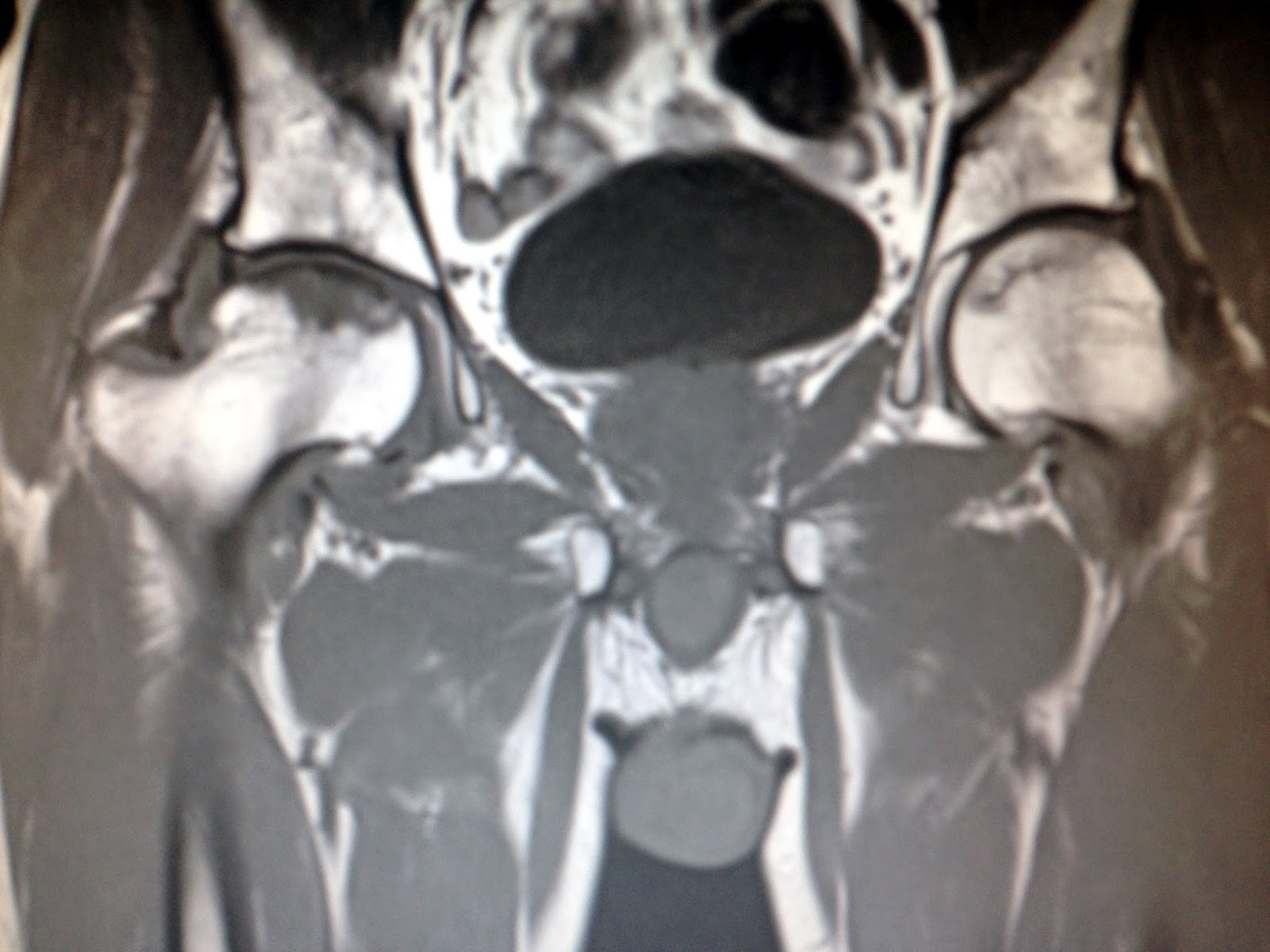
CT Scan abdomen: A mass of 11 x 9 cm with thicken enhancing walls was seen in pelvis.


But diagnosis of gossypiboma was made and at laparotomy: a surgical sponge (18x22 cm) with adjacent inflammatory tissue and pus were removed successfully.


DISCUSSION:
A diagnosis of gossipiboma pre-op seems to be very difficult that always need skill and experience. Because of  imaging findings of gossypiboma are nonspecific and complexe so the right diagnosis in pre-op is still acchived about 1/3 of cases in literature.
But whenever an unknown mass into abdomen with exist surgical scare that should dissolve it may be a gossypiboma or not.


Wednesday 30 April 2014

CASE 252: NECK PAIN in LEMIERRE'S SYNDROME, Dr PHAN THANH HẢI, MEDIC MEDICAL CENTER, HCMC, VIETNAM.


Girl 13 yo, for one week sore throat and  fever, being  treated with antibiotics, now pain at right neck.
Ultrasound at the neck detected on right side some lymph nodes 2-3 cm at posterior SCM. And internal jugular vein dilated, big diameter 2, 2 cm black lumen, no flow,  cannot compressible (see US pictures of CCA and IJV on right side).


Meanwhile, on  the left neck,  there were normal flow of CCA and IJV ( see video clip).

CDI OF CCA AND IJV on R NECK


VIDEO 2: CROSSECTIONAL


What is your emergent thingking? What is the lab you must execute evaluation ?.


Blood tests: WBC= 15k, neutro 40%, CRP=20mg/l  very high,   D-Dimer=500ng/ml. Bacteriology  blood culture is on the way.
Based on CLINICAL INFECTION and  LAB REPORTS, and ULTRASOUND  IMAGES of THROMBOSIS OF IJV, suggestion  of  LEMIERRE SYNDROME.
Urgent treatment with  IV ANTIBIOTICS and ANTICOAGULATION  DRUGS.


REF..HISTORY OF PROF  ANDRE ALFRED LEMIERRE.


After one week treated with antibiotics and anticoagulant, the flow intra right IJV returned.
See CDI and MSCTA images.


Saturday 26 April 2014

CASE 251: PAIN at RIGHT HIP, Dr PHAN THANH HẢI, MEDIC MEDICAL CENTER, HCMC, VIETNAM


Man 52 yo with history of pain at his right hip  joint 2 years prior, recently the pain is getting more severe, cannot walking (photo).


Plain XRay film of the pelvis looked like normal right and left hip joints.


Upon ultrasound  the right hip joint showed  widering of  the hip joint space with fluid collection, and abnormal echostructure of  the head of femoral bone (see 3 ultrasound pictures at right hip).  


Ultrasound examination of the right hip report was abnormal  in suggesting  arthrosis of right hip joint).
MRI of  the hip joint showed that right femoral head  in necrosis and hydarthrosis, and  small change also at left hip joint.


MRI report is aseptic necrosis of femoral  head  on right and left sides. 


Anatomy of vascular supply of femoral  head : 2  anterior  and posterior  pictures.