Total Pageviews

Saturday 26 April 2014

CASE 251: PAIN at RIGHT HIP, Dr PHAN THANH HẢI, MEDIC MEDICAL CENTER, HCMC, VIETNAM


Man 52 yo with history of pain at his right hip  joint 2 years prior, recently the pain is getting more severe, cannot walking (photo).


Plain XRay film of the pelvis looked like normal right and left hip joints.


Upon ultrasound  the right hip joint showed  widering of  the hip joint space with fluid collection, and abnormal echostructure of  the head of femoral bone (see 3 ultrasound pictures at right hip).  


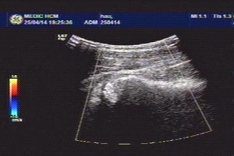
Ultrasound examination of the right hip report was abnormal  in suggesting  arthrosis of right hip joint).
MRI of  the hip joint showed that right femoral head  in necrosis and hydarthrosis, and  small change also at left hip joint.


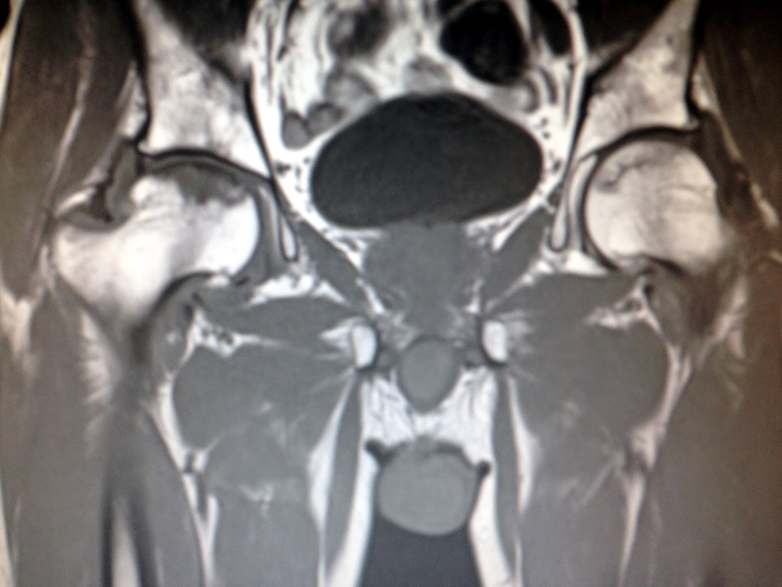
MRI report is aseptic necrosis of femoral  head  on right and left sides. 


Anatomy of vascular supply of femoral  head : 2  anterior  and posterior  pictures.


Monday 21 April 2014

CASE 250: LOST of DENTAL PROSTHESIS, Dr PHAN THANH HẢI, MEDIC MEDICAL CENTER, HCMC, VIETNAM


Man 68 yo (photo) after  drinking a lot of  alcohol, he detected  lost of  dental prosthesis  and dysphasia and apnea,  like  cardiac  ischemia.


 He  came seeing cardiologist for dissolve his cardiac problem, but it was not getting better (echocardio image).


Chest XRay on PA  was normal, but  on LATERAL VIEW  there was something  in retrocardiac space (chest  XRay).


MSCT scan detected  foreign body in the midlle of his esophagus ( see 4 CT  pictures).


Endoscopy  detected at the middle esophagus  one dental  prosthesis made stuck  this site (see photo).


Treatment   emergency   at ENT  hospital. Endoscopy  removed  this  dental  prosthesis   that had lost 3 weeks  before.  Nasogastric  feeding, and wait for  progress. 


See  photo of dental prosthesis.


Sunday 20 April 2014

CASE 249: RLAQ MASS, Dr PHAN THANH HẢI, MEDIC MEDICAL CENTER, HCMC, VIETNAM


Woman 56 yo, pain in RLAQ for 2 weeks, fever and GI tract trouble with diarrhea was treated  by  antibiotics.
Ultrasound detected one mass at RLAQ, suspected appendicular abscess or coecum tumor (see 4 US pictures).


Blood test  WBC= 17K with 17% neutro.
MSCT with CE  shows  this mass  being wall-off  by  instestine,  central part is  liquide as pus collection (see 3 CT pictures).


What is your suggestion of diagnosis and what is  the another test needed for make sure diagnosis?.
Open operation  detected  this mass at  RLAQ  is abscess due to appendicular necrosis.


DISCUSSION: TRIPLE TEST  of appendicitis is blood tests  for diagnosis of acute or   appendicula abscess including 3 criteria: WBC, NEUTRO%  and CRP.

REFERENCE:


Monday 14 April 2014

CASE 248: GASTRIC WALL TUMOR, Dr PHAN THANH HẢI, MEDIC MEDICAL CENTER, HCMC, VIETNAM


Woman 75 yo vomiting, endoscopy detected  an extragastric fundus tumor (see  pictures).


Ultrasound of abdomen showed one hypoechoic mass with size of 4 cm, well-bordered at the hilus of  spleen ( see 2 ultrasound pictures).


MSCT with CE  found out this mass bending the wall of great curvature of stomach, very slow CE enhancement (see 3 CT pictures).


Blood tests of all  markers are normal.
What is your suggestion of diagnosis?
Open surgery removed the tumor easily. It grew from gastric fundus wall, its structure was hard.


MICROSCOPIC  REPORT  WITH  IMMUNOHISTO CHEMISTRY  is  GIST OF  STOMACH WALL.

REFERENCE: