Total Pageviews

Tuesday 5 March 2013

CASE 170: A BREAST TUMOR, Dr PHAN THANH HẢI, MEDIC, MEDICAL CENTER, HCMC, VIETNAM

Woman 77 yo, by herself detected one lump at her left breast.
Ultrasound  examination: this mass was at  section  of 10 hr  of left breast, size  arround 2 cm ( B mode US picture). 

It was  hypoechoic and  irregular  border, with very strong shadowing (image 2 and 3), and on CDI, hypervascular and  very high PI.


On PDI again, this tumor was hypervascular; axillary scan no detected nodes.


Ultrasound first  suggests breast cancer, next step is mammography or  MRI.
 
THIS PATIENT  REFUSED  TO DO MAMMOGRAPHY AND MRI BECAUSE  THE FIRST TECHNIQUE  WAS PAINFUL  AND THE SECOND ONE MADE  CLAUSTROPHOBIA FOR  HER LONG TIME AGO.
MSCT  IS CHOSEN FOR STAGING  THIS CASE. (SEE  3 CT SLICES )


MSCT non CE  showed that tumor  was small size of  1.8 cm, spiculate hypercalcification and detected no  lymphatic nodes of axillary or retrosternum, it was staging I.
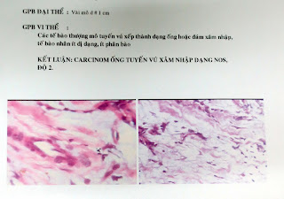
Biopsy was done and report was breast cancer type NOS.


Tuesday 19 February 2013

CASE 169: CBD MASS, Dr PHAN THANH HẢI, MEDIC MEDICAL CENTER, HCMC, VIETNAM

MAN 50 YO, ONE MONTH AGO, PAIN AT RUQ AREA,  FEVER AND JAUNDICE PROGRESSING. ULTRASOUND AT FIRST DETECTED THE GALL BLADDER TOO BIG, NO STONE AND DILATED CBD with DIAMETER OF  2.6 CM,  WITH  PENCIL SIGN  AT ODDI AMPULA. [SEE 3 ULTRASOUND  IMAGES].


MDCT  OF ABDOMEN WITH CE  SHOWED  ODDI AMPULA  HAVING  A MASS  WITH CE  (SEE  2  CT IMAGES).


ERCP was done and  detected  ampular  tumor. Biopsy was performed  and  left a stent for biliary decompression.
Microscopic  report  from  biopsy is adenocarcimoma of Vater ampulla tumor.

Reference:  January 1993 Buck and Elsayed, RadioGraphics.


Monday 11 February 2013

CASE 168: NOT HAVING GALLBLADDER, Dr PHAN THANH HẢI, MEDIC MEDICAL CENTER, HCMC, VIETNAM

Man 48 yo in hospital admission by fever and jaundice progressing. Physical examination: pain at right subcostal area, past history was known diabetes and gallbladder stone. Plain abdominal XRay film standing showed ilius status.


Ultrasound of abdomen revealed small fluid collection at the border of liver, and CANNOT FIND OUT THE GALL BLADDER BUT DETECTED ONE  HYPERECHOIC MASS ADHERED TO LOWER BORDER OF LIVER. THE CBD WAS NOT IN DILATATION.


Blood tests with elevated WBC of 16K (90% neutrophil).

MDCT non CE found that the gallbladder without stone nor fluid into gallbladder.


What is your explanation of the ultrasound images for this gallbladder?


Based on clinical  status:  fever, jaundice, pain at right subcostal area, and imaging modalities (abdomen plain film, ultrasound  and MDCT) with  blood tests, the  diagnosis was acute cholecystitis lead to gallbladder empyema. The IV antibiotic resulted clinically good response in medical treatment.

Reference: 


Tuesday 5 February 2013

CASE 167:THYROID NODULE in a GIRL, Dr PHAN THANH HAI, MEDIC MEDICAL CENTER, HCMC, VIETNAM

An 11 yo girl was detected her right neck mass by her mother.
In clinical examination at Children Hospital  she was suspected a thyroglossal cyst, but on ultrasound scan it was a solid tumor with size of 2cm, central cystic necrosis and hypervascular intramass.


 Blood tests were normal and thyroid isotopic scan was a cold nodule.


FNAC WAS DONE AND CYTOLOGY  REPORT  WAS  COLLOIDAL GOITER.


DISCUSSION: ULTRASOUND  WITH  COLOR DOPPLER IS  THE  FIRST CHOICE  FOR  THYROID  EVALUATION, IN COMPARISON TO  ISOTOP SCINTIGRAPHY WHICH IS  NOT IN ROUTINE USAGE  FOR  PEDIATRIC PATIENT.

Friday 25 January 2013

CASE 166: SWELLING of DISTAL THIGH, Dr PHAN THANH HẢI, MEDIC MEDICAL CENTER, HCMC, VIETNAM


Man 19 yo,  pain and swelling at distal right femur one month ago (see photo).
 
 
Ultrasound examination this thigh with linear and convex probe showed the mass was fro periosteum extending to muscle and destruction the bone  like sunray image.


Front and lateral X-ray films this mass were not clear, but MSCT revealed the mass being from periosteum and erosion the cortex of the femur bone with periosteal reaction both 2 sides.


Biopsy was done,   microscopic report was DISTAL FEMUR TUMOR. Report of biopsy of this tumor is OSTEOSARCOMA. THE TREATMENT CHOICE IS AMPUTATION THE LEG OR REGIONAL CHEMOTHERAPY.

Thursday 17 January 2013

CASE 165: A SUBCUTANEOUS WALL MASS, Dr PHAN THANH HẢI, MEDIC MEDICAL CENTER, HCMC, VIETNAM

WOMAN 38YO, ONE YEAR AGO SHE DETECTED A  SUB CUTANEOUS MASS AT RIGHT SUPERIOR QUADRANT, MORE AND MORE HARDENING AND CHANGING SKIN COLOR ABOVE IT.(SEE PHOTO)


ULTRASOUND SCANS THIS INTRAABDOMINAL WALL MASS, SIZE OF 7 CM, MULTISEPTATION HYPOECHOIC LIKE CYST, UNCOMPRESSIBLE. SEE 4 ULTRASOUND IMAGES.

MSCT WITH CE=IT IS A ROUND SUBCUTANEOUS TUMOR  NO INVASION TO MUSCLE. SEE 3 CT PICTURES.


MICROSCOPIC  REPORT  THIS CASE IS  DERMATOFIBROSARCOMA PROTUBERANS, IT IS LOW GRADE  SARCOMA.
Ref.:


Sunday 13 January 2013

CASE 164: ASCITES due to MALIGNANT MESOTHELIOMA, Dr PHAN THANH HẢI,MEDIC MEDICAL CENTER, HCMC. VIETNAM


Man 64 yo, for a long time abuse of alcohol, he himself detected abdomen distention.

At MEDIC,  ultrasound of abdomen and MSCT were done for him.


Blood tests:  normal liver function tests, normal renal function, protide total 6,08 g/L; CEA =195 UI/mL. Other cancer markers: CA 19-9, PSA, AFP, beta2 MICROGLOBULINE =1651 (N=2000). ANA negative.
ASCITES FLUID  removed by punction is cloudy yellowish.


Ascites fluid analysis: cytology no malignant cell, 75%  lymphocyte, protid 30,6g/L, sugar 5,3 g/dL, amylase not elevated.
CEA=   650 UI/L;   CA-125 =550 UI/L
ADA = NEGATIVE , PCR TB = NEGATIVE.
Summary =   With blood test and ascites fluid test results , we try to find out cancer by gastroscopy, colonoscopy.
MDCT TOTAL BODY and  MRI abdomen also no detected cancer.


LAPAROSCOPY DETECTED  MANY INTRAPERITONEAL AND PARIETAL SEEDINGS, AND ALSO ON GREAT OMENTUM. THE  COECUM AND  JEJUNUM WERE WALL-OFF AND  ADHERANT  TO THE  ABDOMINAL WALL. LIVER  SUSPECTED NORMAL.  

3 BIOPSIES RESULT  PERITONEAL  CARCINOMATOSIS.

Microscopic report is  mucineous adenocarcinoma  metastasizing to peritoneum.


Discussion: why the diagnosis of  this case  delayed for one month after onset of ascites.
Where is the primary cancer and what is the choice of treatment for for him from now on?

STAINING IMMUNOHISTOLOGICAL RESULT IS A MESOTHELIOMA.

AFTER  SPECIAL EXAMINATION  BY HISTOIMMUNO, THIS  REPORT IS INTRAPERITONEAL MALIGNANT  MESOTHELIOMA.