Total Pageviews

Wednesday 20 April 2016

CASE 374: MALIGNANT PHEOCHROMOCYTOMA and CUSHING SYNDROME, Dr LÊ TUẤN KHUÊ, Dr NGUYỄN MINH THIỀN, Dr PHẠM THẾ ANH, Dr PHAN THANH HẢI, MEDIC MEDICAL CETER, HCMC, VIETNAM


Woman, 20 yo, discovered HTA  in pregnant at 16 weeks
 5-months before this hospitalization: pregnant  in 28ws, sudden obstetrical  seizures in Ca Mau hospital.  Diagnosis of eclampsia / hypertension / 28w pregnant. Treatment: cesarean section.
 After surgery , patients changed  body shape, round, fat face, neck, stretching skin, increasing weight gain, examination findings adrenal gland tumor  in Can Tho General Hospital, then transferred to Binh Dan hospital.
In clinic examination, obesity, Cushing syndrome,  other organs detect no abnormalities. HTA being treated.


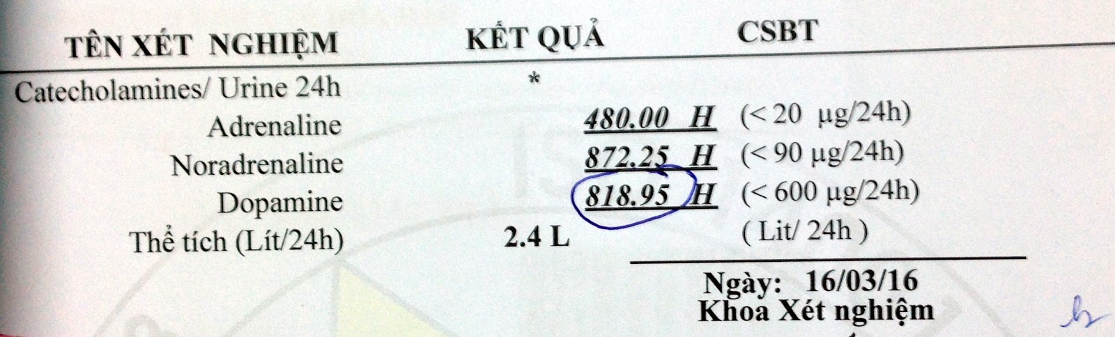
Blood and urine catecholamine increasing, blood and urine cortisol increasing, ACTH reducing.


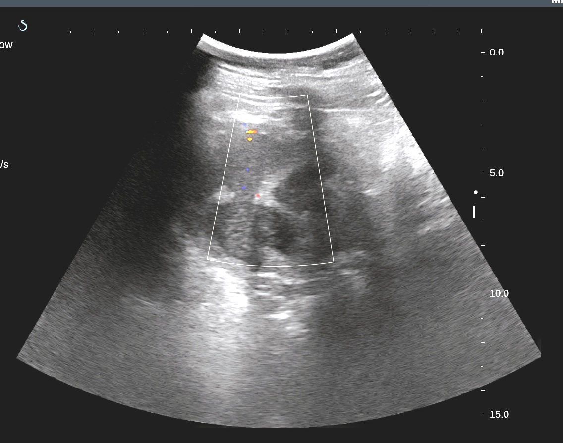
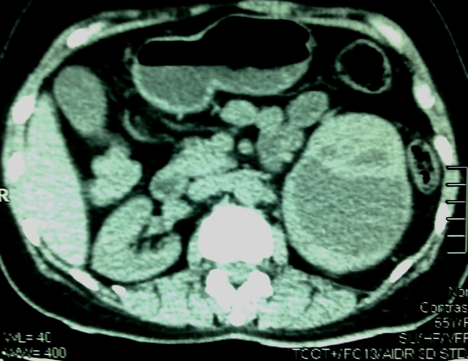
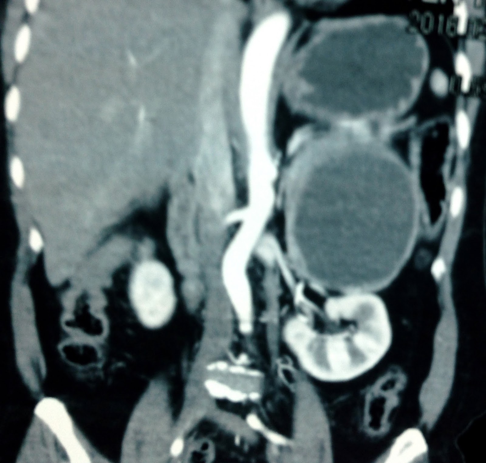
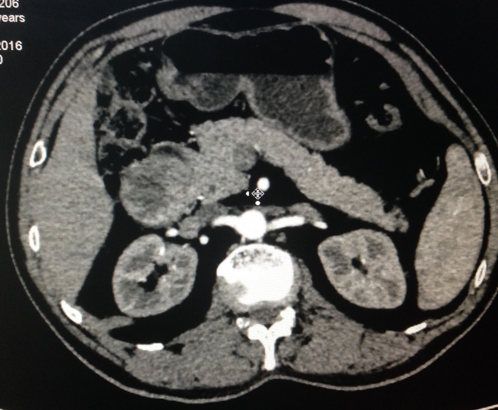
MSCT: right adrenal tumor.

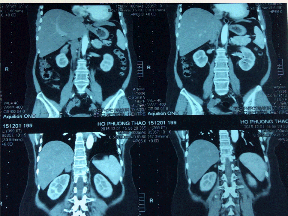
CONCLUSION=  Female patient 20 yo, hospitalized for weight gain and  HTA, Cushing syndrome. Reducing of  blood ACTH and metanephrine; catecholamine and cortisol secretion increasing in blood, and urine.
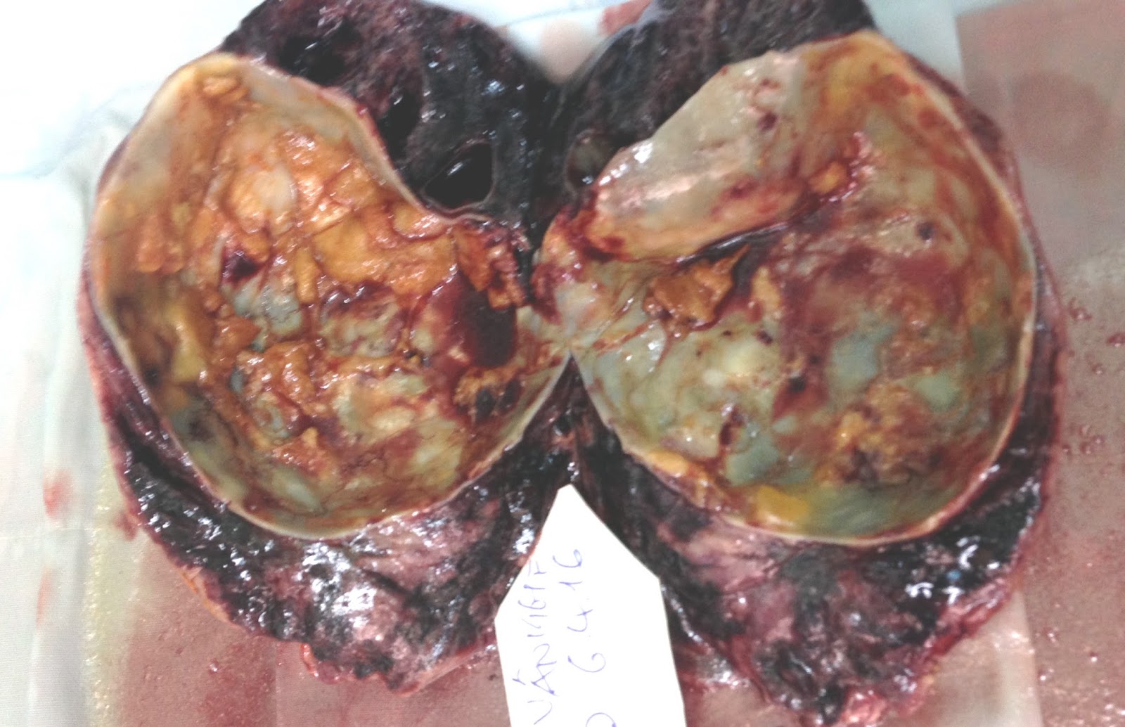
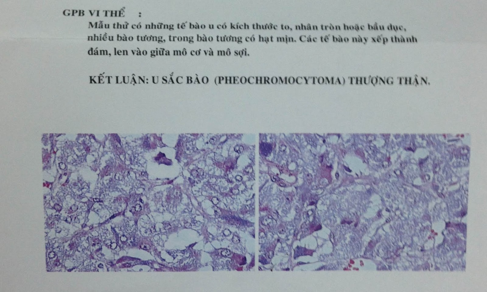
Surgery removed right adrenal tumor. Pathological result is malignant pheochromocytoma.


 Secreted adrenal neoplasms - suppression of axis of  adrenal pituitary.

Sunday 17 April 2016

CASE 373: PHEOCHROMOCYTOMA, Dr PHAN THANH HẢI, MEDIC MEDICAL CENTER, HCMC, VIETNAM


Woman 55 yo with blood pressure rise  crisis for 5 years,  max 20/10cm Hg, medical treatment cannot control in stable. Ultrasound of abdomen detected one round mass  at  left adrenal area, ..size of 8cm  well bordered, cystic with septation ( US 1, US 2).


MSCT with CE=  CT1: sagittal plan this mass at  adrenal fossa  deplaced  left kidney. CT2:  normal vascular  supply to kidney . CT3: crossed section of.this mass inhomogeneous  in contrast enhancement.


Blood test shows very high  catecolamine of 24 hours in urine  and metanephrine in plasma is 1521.53/mL  (normal <90 ng/mL).

,

Diagnosis of this case is pheochromocytoma which  based on clinical,  ultrasound, CT and  blood test.
Operation removed the tumor (see macro 1,2).

MICROSCOPIC REPORT  IS  PHEOCHROMOCYTOMA.


POST OP   BLOOD PRESSURE IS STABLE.

Reference:


Friday 8 April 2016

CASE 372: PORT-SITE METASTASIS, Dr PHAN THANH HẢI, MEDIC MEDICAL CENTER, HCMC, VIETNAM


W
oman 50yo,  6 months before  had been laparohysterectomy by endometrium carcinoma.  One  week  ago  she detected  pain  at  RLQ area at the  site of puncture for operation before (photo). 


Ultrasound scan  with  curve  probe for  this mass is hypoechoic structure in the wall of abdomen (US 1).


US 2  CDI   vascular supply from  the muscle arround


and
US 3  scanning with  linear probe = this mass is  in abdominal wall.


MSCT  with CE of this mass is enhanced with contrast  and  located in abdominal wall (CT 1, CT2).


Core biopsy of  this mass with microscopic report is adenocarcinoma


in metastasis on the site which was  laparoscopic tap of endoscopic operation.

DISCUSSION...IT IS METASTASIS AT  ABDOMINAL WALL  AFTER  LAPARO-PORT -SITE. AFTER  6 MONTHS THIS TUMOR  IS GETTING GROWTH VERY FAST.

REF PDF.

Saturday 2 April 2016

CASE 371: BCCAO (BILATERAL COMMON CAROTID ARTERY OCCLUSION) Dr PHAN THANH HẢI. MEDIC MEDICAL CENTER, HCMC, VIETNAM


Man 38yo, high BP with headache,


 psoriasis only on hands (see foto of hands),


 in  routine ultrasound screening of  vascular neck detected  bilateral common carotid  stenosis completely with dilated  vertebral arteries  both 2 sites (see  US 1,2= R-L.CCA, US 3-4=R-L/ I+ECA, US 6-7= R-L/ Vert.A).


MSCT Angio showed that completely obstruction of right and left CCA.


Patient has not loss of vision or any neurological symptom.

Blood test on DDMERE [D-DIMER] was in normal level of 270 ng/mL.

Discussion: Do you see the BCCAO case  looked like this ? What is the cause of disease?
Reference: One case of BCCAO.


Tuesday 29 March 2016

CASE 370:PLEURAL EFFUSION, Dr PHAN THANH HẢI, MEDIC MEDICAL CENTER, HCMC, VIETNAM


Man 21 yo  fever and  cough , thorax pain in breathing
Chest XRays suspection  of pleural effusion.


CT scan  with CE of the chest:
CT1: cross- section=.pleural effusion both 2 sites.
CT2 .frontal section.
CT3:   frontal section in anterior  mediastium detected  one mass at retrosternum.


Blood test of WBC not raising.
Pleural tap  removed  yellowish liquid, but  analysis was lower ADA level.


TRANS THORACIC ULTRASOUND AT SUPRASTERNUM DETECTED ONE   RETROSTERNUM MASS, LOOKED LIKE LYMPH NODE (US1).


PLEURO-ENDOSCOPY FOR EXPLORATION= NON DETECTED  INTRA PLEURAL LESION AND REMOVED THIS  RETROSTERNUM MASS( MACRO)
MICROSCOPIC REPORT  THIS MASS IS NORMAL TISSUE OF THYMUS GLAND.


Tuesday 22 March 2016

CASE 369: DUODENUM TUMOR, Dr PHAN THANH HẢI, MEDIC MEDICAL CENTER, HCMC, VIETNAM


Man 60 y.o., health check-up  by ultrasound of  abdomen detected  one mass  nearby gallbladder, size around 3-4 cm.
US 1: this mass  near  gallbladder and  duodenal bulb.

US 2: no  relation to  liver hilus.

  
Endoscopy report that  lumen of duodenum D2 is compressed by external mass (see endoscopy picture).


MSCT  of abdomen with CE
CT1:  this mass is  from  duodenum D2.

CT2 :  patient in rotation  for good vision of pyloris and duodenum mass at D2.


CT3: sagittal section of this mass is CE enhanced.


Preoperative diagnosis is duodenum wall tumor which was  suggested to GIST.

Operation  resection of  tumor and  performed gastro-enterostomy.


Microscopic report of this tumor is GIST.


Reference:
Duodenal gastrointestinal stromal tumor
http://www.sciencedirect.com/science/article/pii/S1743919112007649