Woman 30 yo with total colectomy by colon
poliposis for 2 years ; one month ago she detected
ascites
unknown
origine at MEDIC [19,
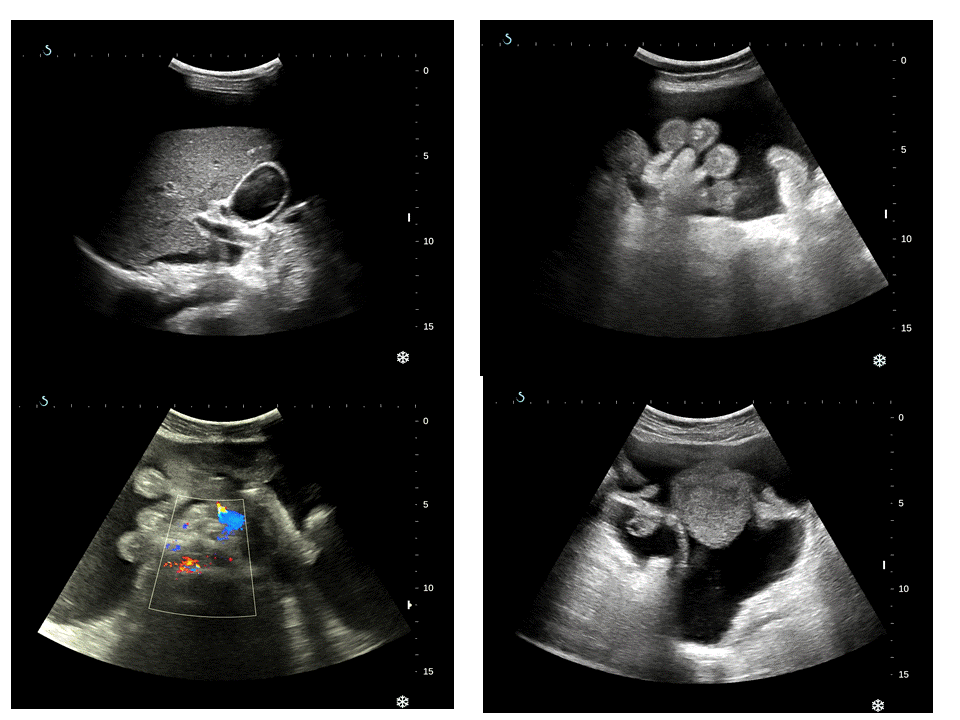
April]. Ultrasound
showed that high volume ascites, normal liver
and.kidney (see 4 pictures ultrasound).
CT of abdomen
with CE also cannot detected the cause of ascites; she underwent laparoscopic biopsy of peritoneum and report was non specific chronic inflamation.
One week later [ 25,April, 2016] she got acute abdomen pain..and came to MEDIC again.
CT of abdomen
with CE detected left kidney hydronephrosis 2nd degree and one
mass of 5 cm in retroperitoneum near abdominal aorta bifurcation obstructed left ureter loodk like urinoma (see CT 1 and ultrasound images of this mass(see US
2).


Abdominal
tap removed pink ascites fluid and analysis report= ADA negative, high protein, normal amylase, urea= 36.04mg/dL, creatinine
3.2 mg/mL (normal
<1mg/ml).

Summary of this
case: ascites with CT and ultrasound detected urinoma and high creatinin in ascites that proved an uremic ascites.
Reference:

Reference:
No comments :
Post a Comment