Woman 33 yo PARA 1011 underwent C-section 3 years [ 2013] now ultrasound detected in 6 week pregnancy. But she would like to evacuate the embryonic sac by curettage . Then 3 weeks later she get pain at her pelvis.
Ultrasound again
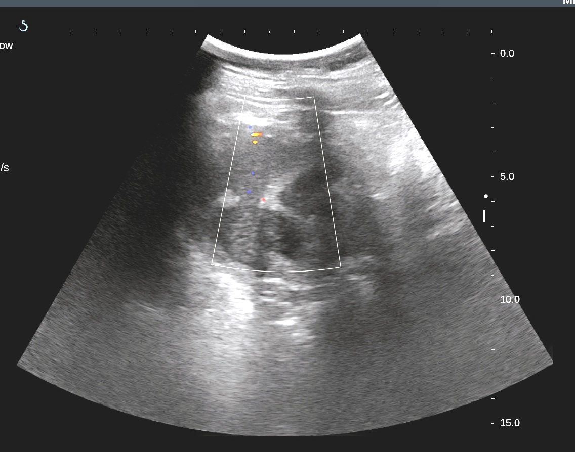
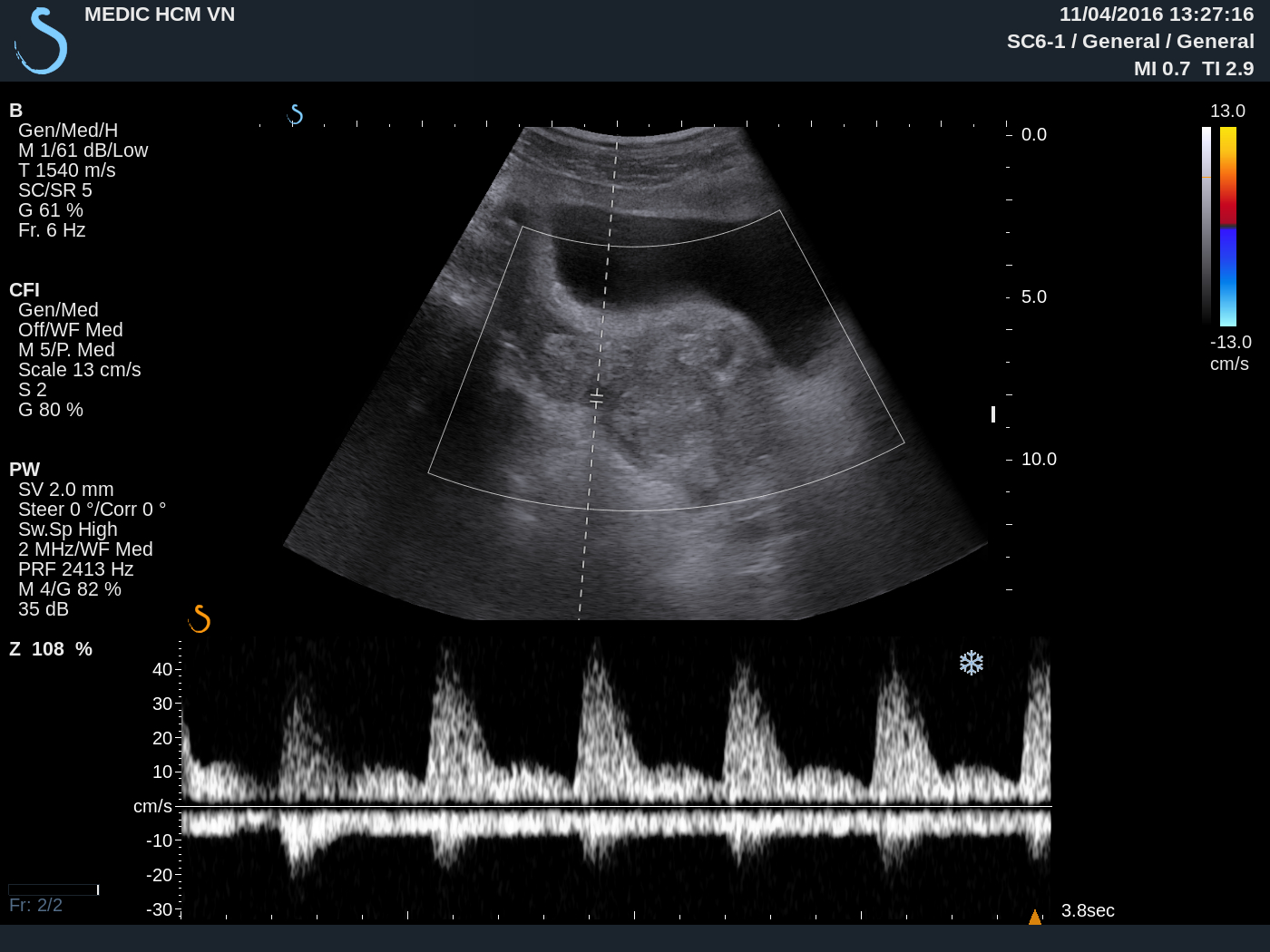
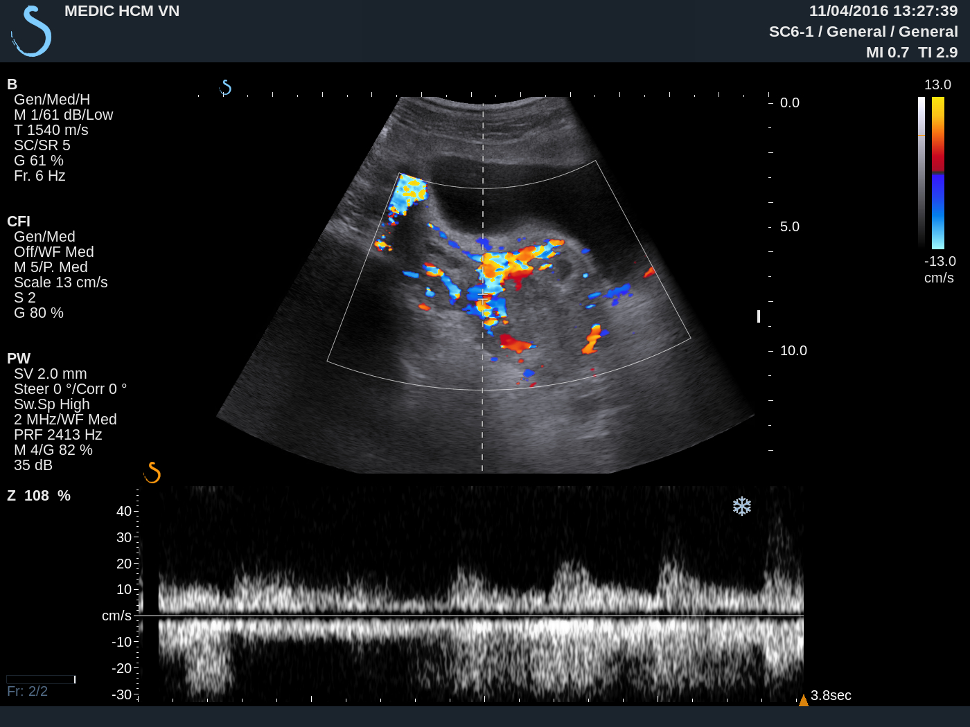
detected one hypervascular mass at the neck of uterus ( see 3 US
images).

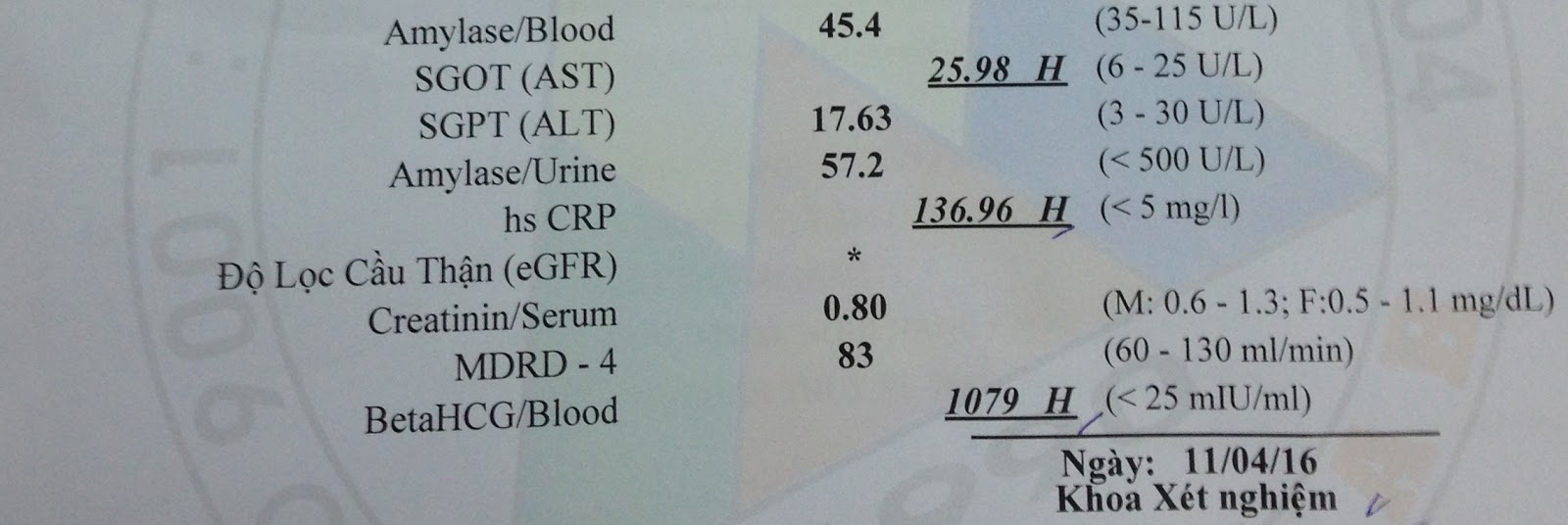
And beta HCG of blood
test is high.
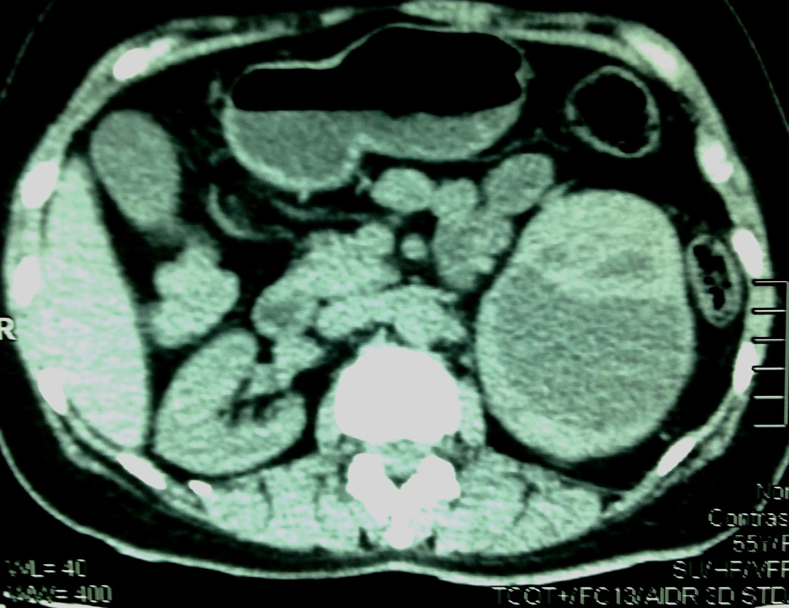
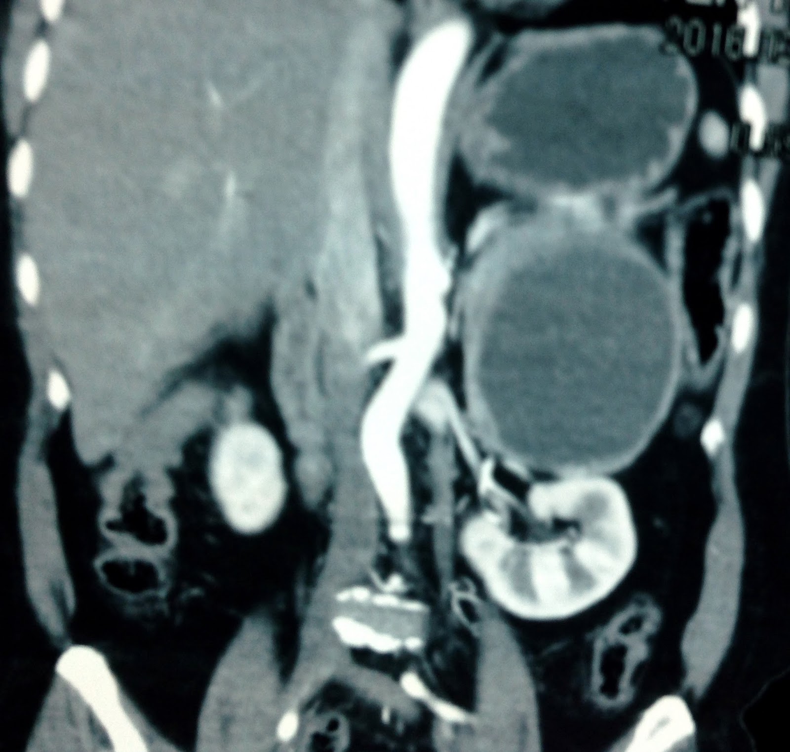
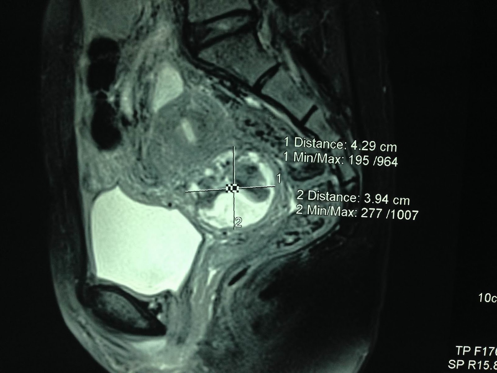
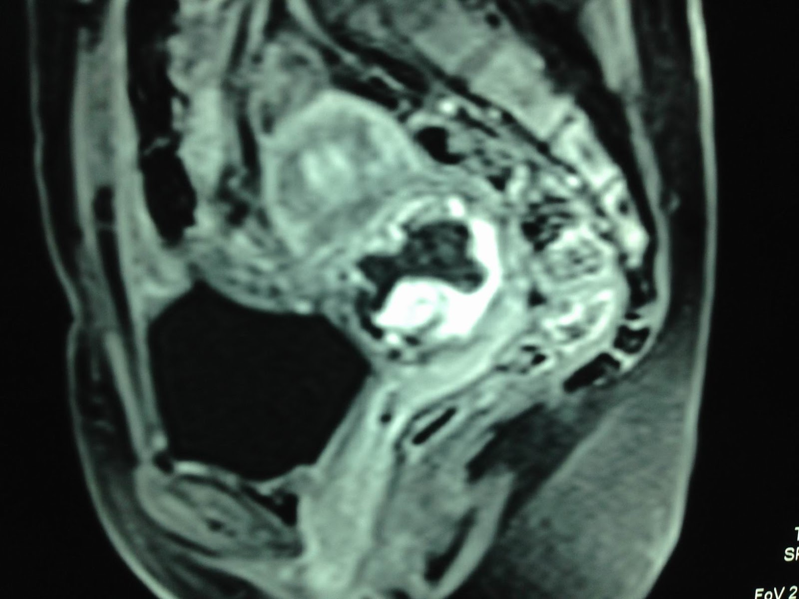
MRI
uterus
of this mass suspected gestation at neck uterus
in the scar of cesarian section before.

Open operation for
hysterectomy confirmed cervical pregnancy in C-section scar.