
Woman 62 yo, cough and dyspnea, weakness of left side of her
body 2 weeks ago.
Chest XRay
first.( see pleural effusion at right lung).

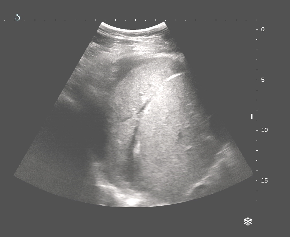
Ultrasound of thorax:
US1=liver normal with mass
at lower portion of right lung

US 2=liver and right
lung looked like liver structure (hepatization).
US 3= scan at right
thorax: pleural effusion and lung solid mass.

US 4= with 10MHz
linear probe looking of visceral layer of pleural membrane having
irregular nodular mass.
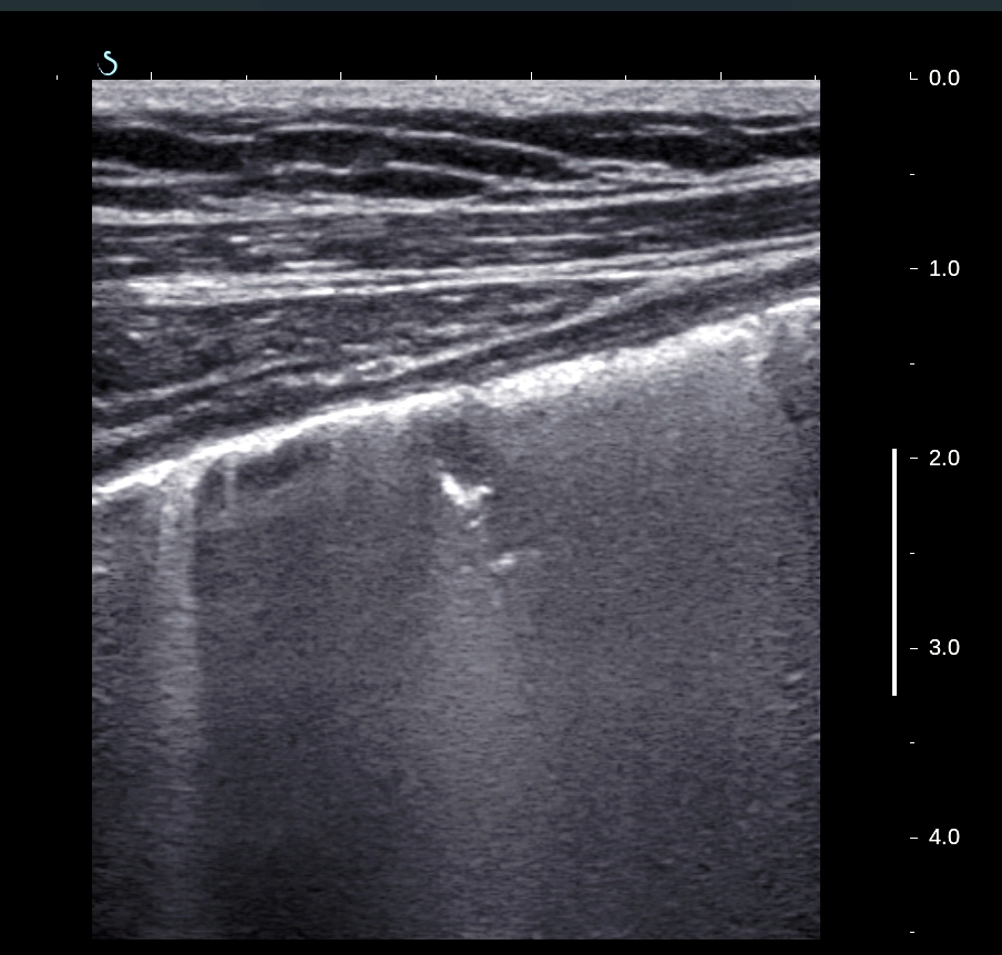
US 5 = this lung mass is hard like liver.

US 6= very low vascular
supplying.

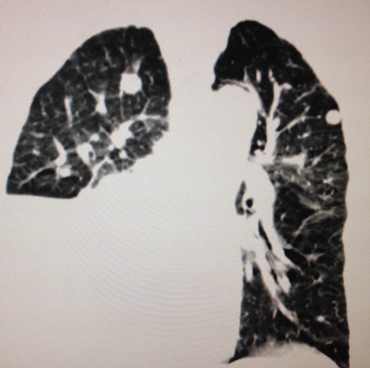
CT scan of lung non
CE.: CT1=cross section, CT2 = frontal view, CT 3= many nodular metastasis at right
and left lung.


CT4= brain scan
with suggestion of metastasis at right brain..
Punction of pleural
space removing yellow fluid ( foto).

Analysis of fluid =
ADA very low, ruling out lung tuberculosis.
Do you thing
this case is lung cancer metastasis to the brain?
REFERENCE:
Ultrasound detection of Lung Hepatization
REFERENCE:
Ultrasound detection of Lung Hepatization
No comments :
Post a Comment