FOR PICTURES PLS CONNECT TO 3G / DOWNLOAD THE LINK
case-322-ovary-twisting-
case-322-ovary-twisting-
WOMAN 36 YO, 3 DAYS AGO PAIN AT SUPRAPUBIS, AND
POLYKYURIA.
ULTRASOUND OF PELVIS WAS NORMAL.
PAIN CONTINUING AND RISING AND SHE WAS IN ADMISSION OF ONE EMERGENCY HOSPITAL, IN CLINICAL
SUSPECTED RENAL COLICKY PAIN.
ULTRASOUND AGAIN DETECTED ONE SUPRAPUBIS MASS,
SIZE OF10CM, CYSTIC WITH CLOUDY FLUID INSIDE (US 1).

CDI REPORT WAS SMALL UTERUS, CANNOT DETECT VASCULAR
AT RIGHT UTERUS CORNER (US 2).


THIS CYSTIC MASS WITH
MASS INTRA CYST LIKED SEBUM THAT SUSGESTED RIGHT
OVARY TERATOMA IN TORSION.
NO STONE INTRA URINARY SYSTEM.
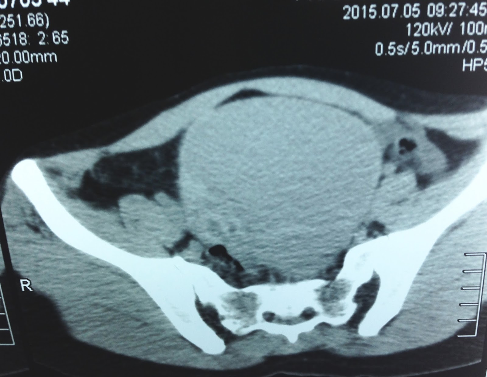
MSCT NON CE PRESENTED OVARY MASS
LOOK LIKED TORSION OF TERATOMA.


EMERGENT OPERATION BY LAPAROTOMY DETECTED THIS MASS BEING AN
OVARIAN CYST BLACK IN COLOR DUE TO
TORSION ( SEE FOTO)


OPEN SURGERY REMOVED RIGHT OVARY MASS
ISCHEMIA AND BLEEDING INSIDE OF A TERATOMA TUMOR.


Discussion : Why the first ultrasound examination was normal? Looking the first picture ultrasound of the uterus that was clear view and the fluid over look liked urinary bladder.
But you can see the small urinary bladder in the lower corner of the first ultrasound picture.
The mistake was due to ultrasound scanning with the urinary bladder not full filling , the ovary cyst was not septation because scanning view in small window.
The ultrasound examination is better view with lateral decubitus position, but in some cases cannot see well in decubitus position.

Pathology report was an ovarian teratoma.

Pathology report was an ovarian teratoma.
No comments :
Post a Comment