Total Pageviews

Friday 27 May 2022

CASE 634: RECTUM CANCER, Dr DƯƠNG NGỌC THÀNH, Dr PHAN THANH HẢI, MEDIC MEDICAL CENTER, HCMC, VIETNAM

 Female patient 55yo loss of weight 7 kg for 4 months with bad feelings of contracting her muscle trying to empty her bowels. There was no blood stool, but existing abnormal uterine bleeding.  Digital rectal exam revealed a rigid, mobile mass at posterior wall that suggested a rectum cancer which took part of 1/3 of lumen of the rectum.

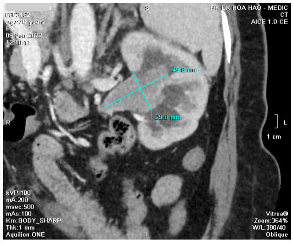
MSCT whole body detected thickening of rectum wall that adhered uterus, captured contrast and blurred fatty tissue around. Results confirmed a rectum cancer invading around with some pelvic lymph nodes.


But ultrasound  and colonoscopy failed  to detect the rectum tumor.


Ultrasound (TVS) only revealed uterine fibroma and cervical polyp. 


It only was in the third time endoscopy detected the rectum cancer. And biopsy results was mucinous adenocarcinoma invaded at rectum.


DISCUSSIONS AND CONCLUSION
  
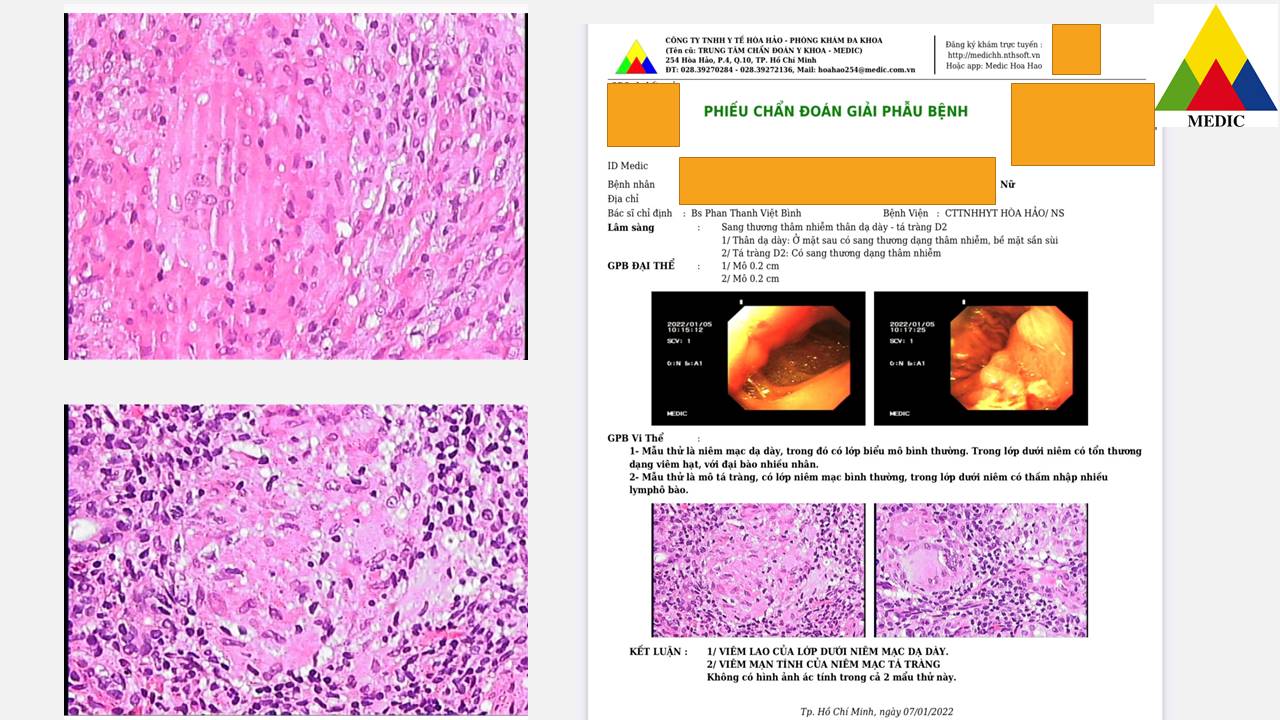
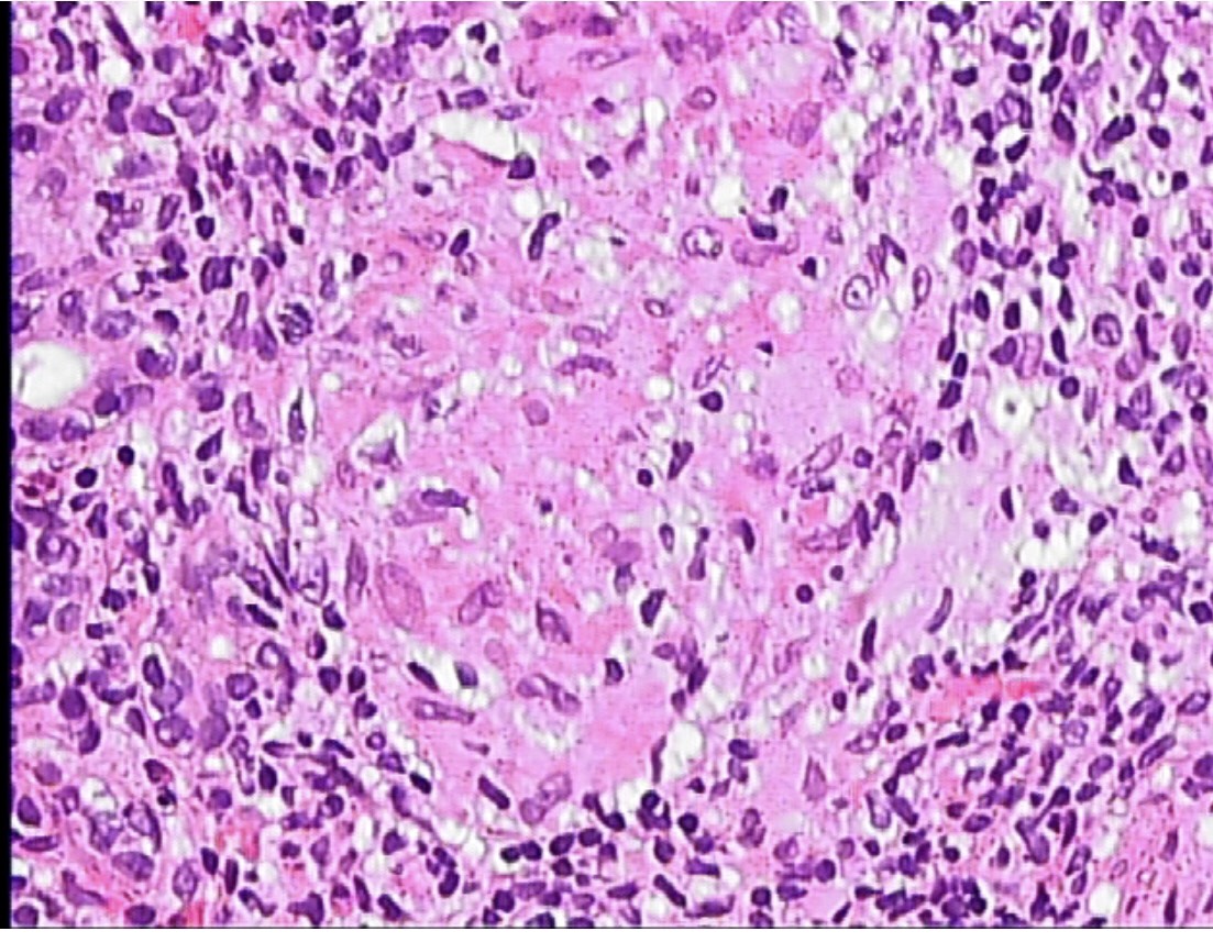
Clinical findings and MSCT  took the clues for diagnosis of the case, but it need  the concordant endoscopic result to make planning of treatment. It was difficult for endoscopy in this case, but at last it existed an evident of anapathology in the third time of endoscopy.

The female patient went through chemotherapy course and later removed rectum tumor in keeping the sphincter muscle of rectum.


Wednesday 25 May 2022

CASE 633: TCC of Kidney, Dr PHAN THANH HẢI, Dr TRẦN THỊ BẢO CHÂU, MEDIC MEDICAL CENTER, HCMC VIETNAM.

Female patient 70yo with dysuria but without hematuria.

Ultrasound detected left kidney hydronephrosis grade 2 as a hyperechoic mass # 47x35mm inside renal pelvis that suggested a transitional cell cancer (TCC).


CT Scan:  Soft tissue mass was in renal pelvis and ureter, d= 30 x 50 mm that highly captured contrast media while left kidney was in poor secretion of contrast. CT confirmed a left TCC.


 It existed red and white blood cells and bacteria in urine analysis.

Endoscopic biopsy results was high malignancy uroendothelial carcinoma  invaded the renal stroma. 


Surgery removed left kidney and ureter. In longitudinal section  of kidney, left pelvic kidney tumor sized # 5cm which was a necrotic vegetative mass while ureter was intact.


Pathological results : Transitional cell carcinoma poorly differentiated invaded parenchymal kidney. Non existed malignant cell in lymph nodes.

Friday 6 May 2022

CASE 632: STOMACH TUBERCULOSIS, Dr PHAN THANH HẢI, Dr PHAN THANH VIỆT BÌNH, MEDIC MEDICAL CENTER, HCMC VIETNAM


Female patient 32 yo, loss of weight #10kg with epigastric pain and nausea for 2 months. 


She herself took gastric drugs for a while but failed so went to Medic for a new examination.

Ultrasound of abdomen at Medic revealed many lymph nodes that were suspected metastatic nodes and mesenteric thickening; stomach walls infiltrated thickening and slight splenomegaly.


Chest X-rays was normally detected.


Gastric endoscopy showed gastric corpus roughly inflammed. Results of biopsy were  inflammed submucosa layer of stomach and chronic inflammation of duodenum suspected due to TB infection.


MSCT confirmed that existed a lot of lymph nodes at hepatic hilus, lesser curvature of stomach, around celiac axis. These nodes maybe belong to TB nodes.


Result of biopsy of intraabdominal lymph nodes was TB inflammed nodes.


Discussions and Conclusions


TB of stomach is still a rare entity, which is about 1-2% of GI tract tuberculosis and in 0.5% of  patients contracted with TB. Usually it is secondary after the pulmonary TB infection.

Our patient is now getting better status, gained more 2 kg of weight while being taken TB drugs for 2 months of 6 month-regimen of therapy.