61 yo female patient went to Medic Center
PA and lateral chest XR revealed of a suspicious big mass of 11x 5cm on L lung base : was that a tumor?
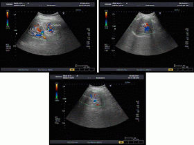
An ultrasound examination was performed to know that mass is solid or cystic nature. To our surprise, a typical structure of a kidney is detected by echography in ectopic situation but difficult to certified it is above or under the L diaphragm.
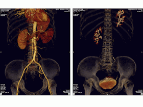
The problem was easily resolved by CT scan with contrast showed nicely the L kidney well vascularized and preserved function herniated through Bochdalek foramen.
So it was an ectopic thoracic kidney and diaphagmatic hernia.


No comments:
Post a Comment