Total Pageviews

Saturday 20 May 2023

CASE 683: A TB CASE: Dr PHAN THANH HẢI, Dr TRẦN THỊ TRÚC PHƯƠNG, MEDIC MEDIC CENTER, HCMC, VIỆT NAM

 A 29 year-old female  patient in a general chek-up for a preparation of a study abroad.

Ultrasound detects lymph nodes in her mesentery in the abdomen and on her neck # 5-25 mm in suspecting TB infiltrating type. Also a slight thickening of wall of colon is noted.


Report of the neck lymph node full biopsy is an TB  infected node.


A colonoendoscopy performs shows findings of ulcerative colitis from the cecum to the transverse colon due to TB infected.


A TB regimen is planned for the patient, and during 2 months of TB management, her status is getting better, that is proven by the clear chest X-Ray and decreasing of lymph node in size and numeration.


Friday 19 May 2023

CASE 682: RIB CHONDROSARCOMA, Dr PHAN THANH HAI, Dr HO CHI TRUNG, MEDIC MEDICAL CENTER, HCMC, VIETNAM.


A 56 male patient with right shoulder pain for a half of month.

Chest X-Ray and ultrasound show a #3.1x2.5 cm right posterior legion of  the 7th rib  with hypoechoic pattern, neovascular signals and moderate hard code on elastography ultrasound.


CT confirms and  suspects malignancy to the # 3.5 cm diameter of rib lesion with erosions of upper border of the 7 th rib which is strongly capturing CE.


Surgery removes the tumor and the histopathologic report is a mesenchymal chondrosarcoma.


It is a rare entity of cancer of  low metastase cartilage in middle aged patient.

Saturday 13 May 2023

CASE 681: PARATHYROID TUMOR, Dr PHAN THANH HẢI, Dr JASMINE THANH XUÂN, Dr HỒ CHÍ TRUNG, Dr LÊ VĂN TÀI, Dr LÊ TUẤN KHUÊ, Dr BÙI BỈNH HUÂN, Dr TRẦN THANH CƯỜNG , MEDIC MEDICAL CENTER, HCMC, VIETNAM.

A 33 year-old male patient with dysuria and nausea.

His 10 year history noted 10 times of long bone fracture, and renal stones and double JJ sonde on the right kidney in hydronephrosis.

Ultrasound detects right hydronephrosis with a sonde JJ, and some renal stones on 2 sides. With experience sonologist herself reveales a left parathyroid tumor next to the lower pole of left lobe of thyroid.


Lab data and X-Rays show a case parathyroid tumor with severe bone complications.


Osteogram: Osteoporosis in severe level.


Surgery removes the left parathyroid tumor  and the PTH level rapidly goes down after removing the tumor.


 For ten years following the initial bone fracture, the patient has experienced many bone problems, including kidney stones. The patient was not able to comprehend the connection between renal stones and bone fractures because the source of the bone fracture and renal stones, which was a parathyroid tumor, had disappeared. 


Wednesday 10 May 2023

CASE 680: CHOLECYSTITIS and GALLSTONE, Dr PHAN THANH HẢI, Dr HỒ KHÁNH ĐỨC, Dr LÊ VĂN TÀI, Dr LÊ THANH TÙNG, MEDIC MEDICAL CENTER, HCMC, VIETNAM.

 A 70 year-old diabetic male patient with an acute abdominal pain for hours enters the emergency room of Medic Center. His prior history are 17 year known gallstone, and denies any RUQ pain or gastrointestinal  troubles.  He had been controlled well his glycemia. He was taken transplant  coronary arterial 2 stents as 2 times of myocardial infarction for over 10 years. His EKG shows a life-rhythmic extrasystolic. WBC: 11.4H, CRP:0.4.

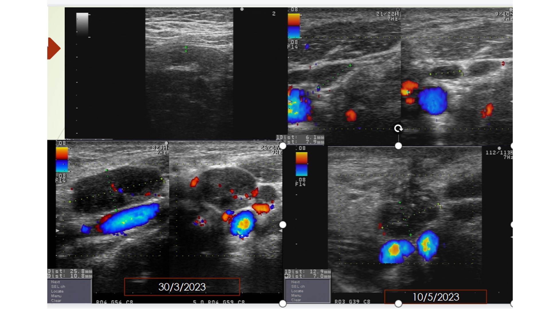
Ultrasound represents a 17 mm stone in a # 98x31 mm gallbladder with thickened wall of GB # 5-7mm. SonoMurphy sign is positive and no fluid exist around the GB.


The cholecystitis pain reduces rapidly with taken diclofenac IM, drotaverin per os after 20 minutes. But a cholecystectomy via endoscopy is planned in regarding his Clopidogrel, diabetic status and the life-rhythmic extrasystolic EKG.

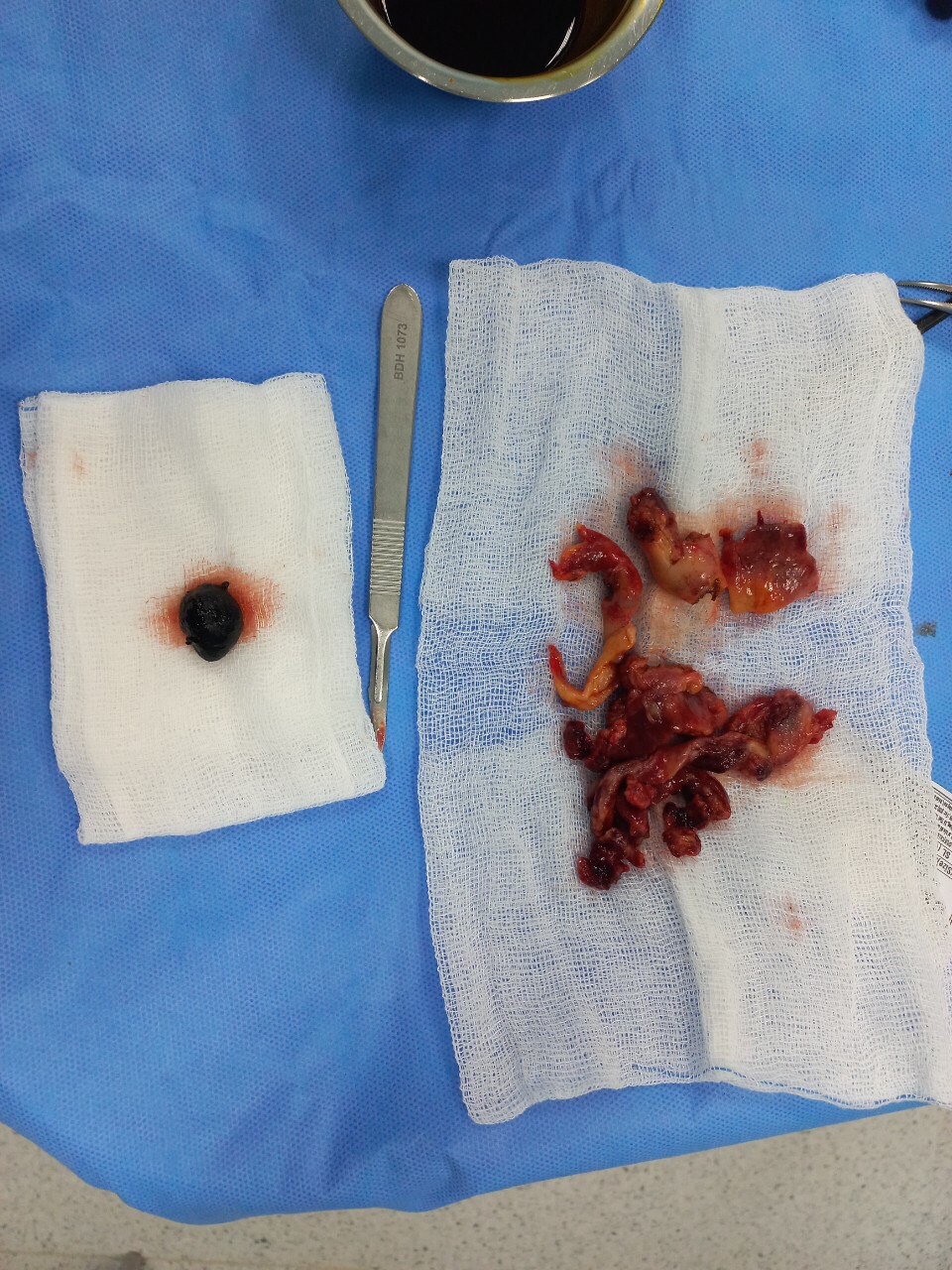
Endoscopic surgery removes a 17 mm pigmented stone within a thicken gallbladder wall. The GB mucosa is partially necrosis and hemorrhage. Thicken GB wall leads endoscopic surgery cut it into small pieces for removing the gallstone and the inflamed gallbladder.

Pigmented gallstone and inflamed gallbladder in small pieces.


Endoscopic view of the  inflamed gallbladder.


Microscopic result is  a necrotic cholecystitis due to gallstone.  The patient remains well and discharged in safety after 2 days in hospital.

In reviewing the gallbladder, the gross specimen of GB shows a cholecystitis image more interesting than the ultrasound view. So it makes alert when facing a painful gallstone than a silent stone of gallbladder.

Furthermore there is a proposal that should think about acute cholecystitis while seeing a gallstone in a RUQ painful patient.

Saturday 15 April 2023

CASE 679: ECTOPIC TESTICULAR TUMOR, Dr PHAN THANH HẢI, Dr BÙI HỒNG LĨNH , MEDIC MEDICAL CENTER, HCMC, VIETNAM.


 A 53 year-old male patient  having HBV infected with hypogastric pain and without his left testis.

Ultrasound detects a # 76x50 mm hypogastric mass, with Doppler signals, and hard code of elastography ultrasound, 23.8 kPA.


MSCT confirms a 75x60x85 mm left ectopic testicular tumor maybe a seminoma in the pelvic region.


Surgery removes the ectopic testicular tumor from left hypogastric region.


The  microscopic result of the ectopic testicular tumor is a seminoma invading its capsule. 

Thursday 13 April 2023

CASE 678: COMPLICATED DIVERTICULITIS, Dr PHAN THANH HẢI, Dr LÊ THỊ THANH THẢO, MEDIC MEDICAL CENTER, HCMC, VIETNAM.

A 55 year-old female diabetic patient suffers from epigatric pain crisii for 4 days and diarrhea.

Ultrasound detects an amount of abdominal free fluid,  two RLQ abscesses, and edema of peritoneum.


MSCT confirms the peritonitis due to 2 abscesses at the right flank of the abdomen.


A right colectomy is performed as perforation of colonic diverticulitis # 2x2 cm of transverse colon and the other one of cecum.

Subhepatic drainage is made to withdraw fluid out and an artificial anus is done.

 And the female patient remains well.


Wednesday 12 April 2023

CASE 677: LUNG TUMOR, Dr PHAN THANH HẢI, Dr VÕ NGUYỄN THỤC QUYÊN, MEDIC MEDICAL CENTER, HCMC, VIETNAM.

A 53 year-old female patient with left thoracic pain for some months. She went through 2  lung tumor (hemangioma) surgeries respectively for 15 years  in K hospital and 4 years (Binh Dan hospital) before.


Pulmonary function report is normal.


Ultrasound notes a round mass  #50x39 mm at the bottom of left lung. It is a cystic lesion, well-limited capsule, and  belongs to the soft code on elastography ultrasound.


MSCT confirms a left pulmonary septated cyst  # 45x29 mm with sludge.


Surgery in the third time (on Tuesday 12, April, 2023) performs for removing the left lung cystic lesion. 

Gross specimen of the left lung tumor.


Microscopic result is malignant hemangiopericytoma.