Total Pageviews

Thursday 5 January 2023

CASE 664: 03 CASES of PROSTATE DISORDERS with LOW VALUES of PSA, Dr PHAN THANH HAI, Dr NGUYEN MINH THIEN, MEDIC MEDICAL CENTER, HCMC, VIETNAM.

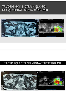
 3 cases of prostate disorders wit low values of PSA= two cases of TB and one case of carcinoma that concludes prostate cancer may happen with any value of PSA [but not only PSA>4 ng/mL]; although value of PSA being considered a guiding lab data for prostate cancer.

Should base on free PSA, DRE, MRI, elasto ultrasound together for screening prostate cancer, but these lab data and diagnostic imaging not stand for a diagnostic of prostate cancer.

CASE 1


CASE 2


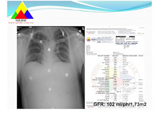
CASE 3:

Chief complaint: only left swollen scrotum.


Friday 23 December 2022

CASE 663: ENDOMETRIOSIS and DIENOGEST, Dr PHAN THANH HAI, Dr NGUYEN THI XUAN HOA, MEDIC MEDICAL CENTER, HCMC, VIETNAM

A 38 year-old female patient, TPAL 1-0-0-1, with sideropenia, dysmenorrhea and menorrhagia that has been diagnosed endometriosis in uterus and right ovary by transvaginal ultrasound  (TVUS) and pelvic MRI.

On MRI, size of endometriosis in uterus # 5×7 centimeter and, in ovary # 1.5 centimeter.


She is managed for iron deficiency, and her endometriosis with Dienogest 2mg one tablet per day. For 5 months, focal lesions in her uterus and right ovary are getting smaller sizes. Dysmenorrhea appears slightly and she is with the new generation of progestin acceptable menorrhagia.


NOTA:


Thursday 22 December 2022

CASE 662: MYOCARDIAL INFARCTUS with ST non Elevated, Dr PHAN THANH HAI, Dr BUI BINH HUAN, Dr VU BICH DOAN TRANG , MEDIC MEDICAL CENTER, HCMC, VIETNAM

 A 68 year-old HTA male patient with right chest pain for 8 hours while holding some water buckets.

Troponin I: 648.6 ng/L, CPK MP: 13.32 ng/mL are elevated, but EKG and cardiosonography have no clue of an acute myocardial infarctus.


About 50% patients of unstable angina  [UA] and NSTE MI have no change of EKG in early time while troponin-I elevated during the first 24 hours.

Based on elevated troponin-I and CPK, the patient is transferred to an emergency hospital after a diagnosis of myocardial infarctus is made.

Saturday 17 December 2022

CASE 661: RCC DETECTED in Check-up ULTRASOUND, Dr PHAN THANH HAI, Dr NGUYEN PHUOC TOAN, MEDIC MEDICAL CENTER, HCMC, VIETNAM

A 45 year-old male employee is detected occasionally a # 25 mm left renal tumor in a check-up by ultrasound in his company. He has not signs of lumbago and hematuria.


MSCT later confirms the 30 mm left kidney tumor at lower pole and anterior face which is thought maybe a RCC.


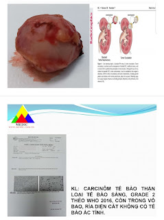
Endoscopic surgery removes the tumor in its capsule by pure enucleation technique.


Result of histopathogy is a clear cell renal carcinoma, grade 2.

Portable ultrasound in control check-up for worker and employee in the companies may detect some cases with subclinical symptom. If it exists any abnormal evident, they will be called going to our center for more informations by other modalities of diagnostic imaging and added lab data. With a final clear diagnosis he/she will be help by an available management.


Thursday 15 December 2022

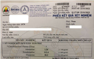
CASE 660: PHEOCHROMOCYTOMA, Dr PHAN THANH HAI, Dr PHAM THE ANH, MEDIC MEDICAL CENTER, HCMC, VIETNAM

A 43 year-old male patient with gastritis and hypertensive crisis, TA=140/100mmHg, P=88b/min.

Ultrasound detects a right adrenal tumor # 41x47milimiter, solid echo poor, less vascularized.


MSCT confirms the right adrenal tumor, with HU density =25 and in late phase of contrast, HU=43.


Surgery removes the tumor carefully in touch due to heart rate and arterial tension going down.


Report of histopathology is a pheocromocytoma of the right adrenal gland.


Saturday 10 December 2022

CASE 659: FIBROSARCOMA of Left SCAPULAR REGION, Dr PHAN THANH HAI, Dr LE VAN THO, Dr HO CHI TRUNG, MEDIC MEDICAL CENTER, HCMC, VIETNAM

A 19 year-old female patient with a huge mass on her left shoulder for 2 years.

Shouder X-ray represents a # 15 centimeter soft tissue mass of left shoulder with invasion left scapulum.                                                  

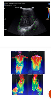
On ultrasound this is a huge solid mass, echo poor, well limited border, poor vascularized from left axilla to posterior shoulder. Thermography notes high temperature in tumoral region.

MSCT shows a soft tissue sarcoma of left shoulder that invades left scapulum. There is an arterial branch from left subclavian artery going to nourrish the tumor.

MRI confirms the soft tissue sarcoma of left shoulder invasive the left scapulum but glenohumeral joint intact.


Surgery removes the tumor and a lower part of left scapulum  keeping of superior part with glenoid fossa of scapulum.

Histopathologic report is fibrosarcoma low malignancy.


Due to COVID19 pandemic, so late the patient goes to hospital in case of tumor with bone metastasis.
Although the surgical management is successful, the patient will still face in high risk of recurrent of  fibrosarcoma and its metastases.


Friday 9 December 2022

CASE 658: CYST of PROSTATE or FISTULA of INTERNAL ILIAC VESSLES, Dr PHAN THANH HAI, Dr LY VAN PHAI, Dr LE THANH LIEM, Dr HO KHANH DUC, MEDIC MEDICAL CENTER, HCMC, VIETNAM


A 67 year-old male patient is detected a small cyst of prostate on the right side by via abdominal ultrasound without any symptom. But on Doppler techniques the real one is a fistula of right internal iliac vessels.
The lesson is a cyst on B-mode may being a dilated vessel on Doppler investigation if sonographer does not apply the Doppler technique to watch a cystic structure.
MSCT and vascular surgery  [vessel collage] proved the  fistula of right internal iliac vessel.


On reexamination, next to the prostate on right side, Doppler ultrasound reveales a # 20x20x24 milimeter aneurysm with arterial low spectral waveform and venous waveform which means a fistula of internal iliac vessels.


MSCT confirms a fistula of the right internal iliac vessels.

An on-line investigation performs with an expert of Binh dan hospital, and this vascular surgeon makes his decision to solve the fistula by collage technique for it, via DSA in his hospital.


The aneurysm of right internal vessel is disappeared on screen while performing of vessel collage technique.


And it exists not any recurrent of right internal iliac fistula on the next 15 days.