Total Pageviews

Friday 9 December 2022

CASE 658: CYST of PROSTATE or FISTULA of INTERNAL ILIAC VESSLES, Dr PHAN THANH HAI, Dr LY VAN PHAI, Dr LE THANH LIEM, Dr HO KHANH DUC, MEDIC MEDICAL CENTER, HCMC, VIETNAM


A 67 year-old male patient is detected a small cyst of prostate on the right side by via abdominal ultrasound without any symptom. But on Doppler techniques the real one is a fistula of right internal iliac vessels.
The lesson is a cyst on B-mode may being a dilated vessel on Doppler investigation if sonographer does not apply the Doppler technique to watch a cystic structure.
MSCT and vascular surgery  [vessel collage] proved the  fistula of right internal iliac vessel.


On reexamination, next to the prostate on right side, Doppler ultrasound reveales a # 20x20x24 milimeter aneurysm with arterial low spectral waveform and venous waveform which means a fistula of internal iliac vessels.


MSCT confirms a fistula of the right internal iliac vessels.

An on-line investigation performs with an expert of Binh dan hospital, and this vascular surgeon makes his decision to solve the fistula by collage technique for it, via DSA in his hospital.


The aneurysm of right internal vessel is disappeared on screen while performing of vessel collage technique.


And it exists not any recurrent of right internal iliac fistula on the next 15 days.


Friday 18 November 2022

CASE 657: MEDIASTINAL ABSCESS, Dr PHAN THANH HAI, Dr PHAN NGUYEN THIEN CHAU, Dr LE HUU LINH, MEDIC MEDICAL CENTER, HCMC, VIETNAM

A 69 year-old male patient enters Medic Center with ten days of fever,  thoracic pain and trouble ingestion. He was managed as gastritis but nothing change.


Blood tests show an infectious syndrome, while EKG, cardiac ultrasound results are in normal limits.


MSCT represents  a # 3 centimeter mass containing air  which is an upper mediastinal abscess with  some calcified foci inside due to a fistula of 1/3 middle part of esophagus.

Surgeon advises immediatly transferring the patient to a surgery hospital.
Gastroendoscopy shows a thickening lesion of middle esophagus and a normal chest X-Ray.


A MSCT is performed to prove the mediastinal abscess, and a bronchoscopy is done to rule out lesion from lung and airways. 

A decision of conservative treatment with antibiotics and a gastrostomy are noted to keep nourrishing the patient which is lasting from now to a half and one month later.


Mediastinal abscess is in recovery phase, reduces its size with calcifications, in two times of re-examination.


This is a mediastinal abscess case due to middle esophagus fistula which is unveiled the cause. 
Clinical clues are fever and thoracic pain and trouble ingestion. The role of MSCT and endogastroscopy are more clearer than chest X-Ray and cardiac ultrasound. Gastrostomy and medical treatment are well enough to help the patient avoiding an unnecessary operation with risks.
And patient remains well and can eating normally by mouth in happiness. 

May the abscess come back?


Thursday 10 November 2022

CASE 656: BOWEL VOLVULUS due to MESENTERIC CYST, Dr PHAN THANH HẢI, Dr TRẦN NGÂN CHÂU, MEDIC MEDICAL CENTER, HCMC, VIETNAM


A 23 year-old male patient with periumbilical pain and left flank pain for 5 days and vomiting. 


Ultrasound detects a cystic mass # 17.9x11.2 centimeter from his navel to pubis, and jejunum dilatation with obstruction sign (washing machine sign). The cystic mass contents fluid and septation with vascular sign on its walls. The cause of bowel obstruction was noted by a non-dilated bowel loop at the mesenteric root with whirpool sign.


There is not  bowel malrotation nor duplication cyst, so the ultrasound findings is bowel volvulus due to a mesenteric cyst.

MSCT confirms bowel volvulus due to a mesenteric cyst later.


Open surgery is done after endoscopic investigation. The cystic mass with yellowish fluid and a part of bowel are removed. Patient remains well post-op.


The histopathological report is a benign cyst with inflammation of the mesentery.
 
Bowel volvulus is still a rare entity especially in young adult.  Mesenteric cyst causes bowel volvulus may happen in emergency room in case of ruling out bowel malrotation, urachal cyst, Meckel diverticulum.


Saturday 22 October 2022

CASE 655: RETROPERITONEAL GANGLIONEUROMA, Dr PHAN THANH HẢI, Dr NGUYỄN KIM HIẾU, MEDIC MEDICAL CENTER, HCMC, VIETNAM

 A 19 year-old female patient with lumbago and periumbilical pain went to Medic Center for ultrasound examination for 10 days.


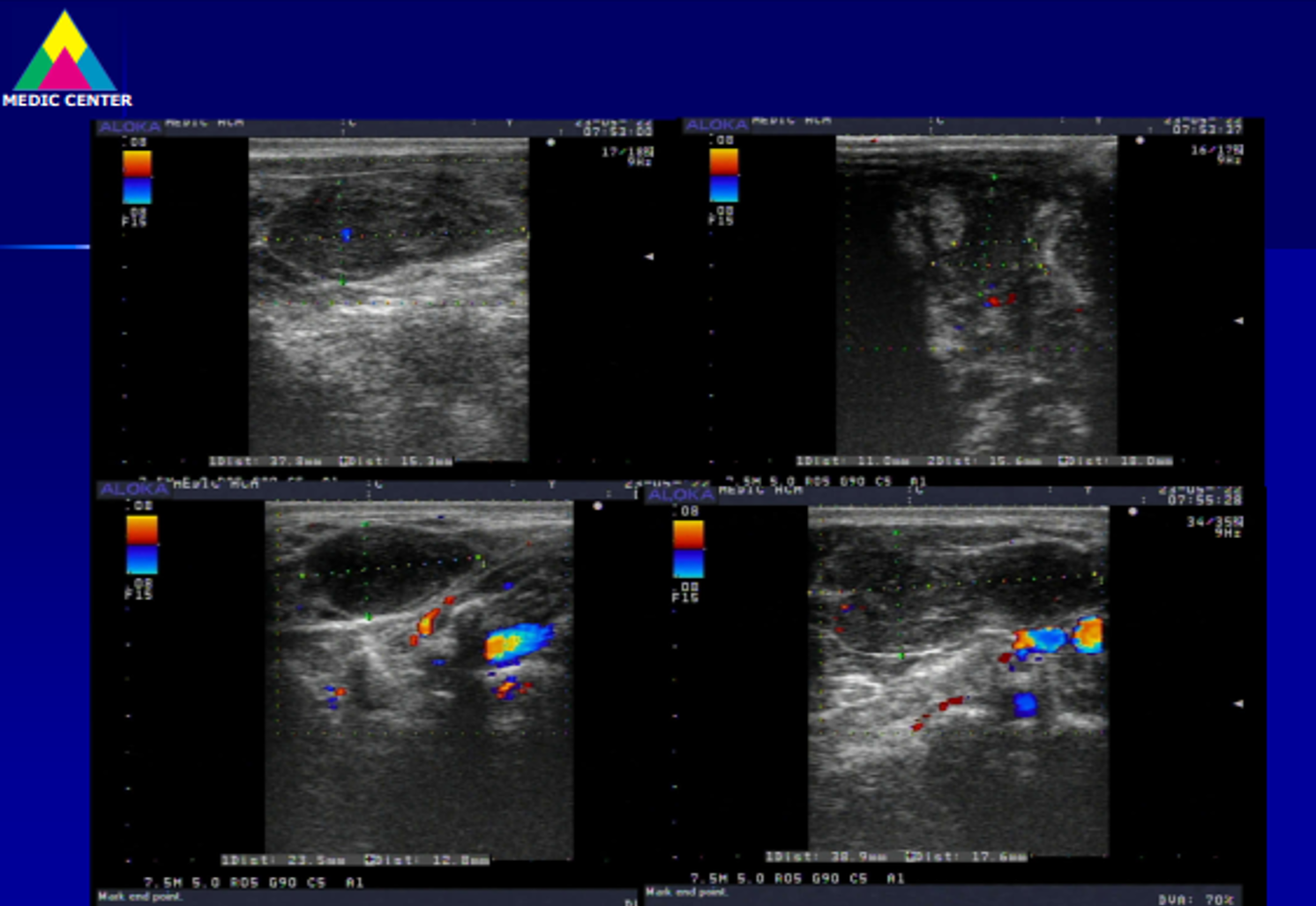
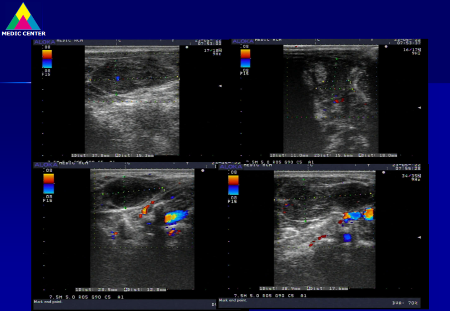
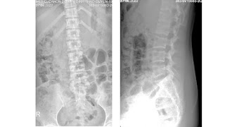
Abdomen ultrasound detects a calcified mass, colorless signal, close by the vertebral column on left side which  is thought a TB abscess or a retroperitoneal tumor. On vertebral X-ray films there are erosions of the vertebral bodies T 11 and T 12.


MSCT confirms a tissue density mass, well limited, with calcifications inside, # 11x17x7 cm, medium contrast captured.  From under the diaphragm the mass compresses left kidney and soft tissues around and erodes vertebral bodies T11, T 12. It may be a retroperitoneal neurogenic tumor.


Surgery was done  after ten days of diagnosing made and post-op result is a retroperitoneal ganglioneuroma.


Now the patient remains well and no need any other treatment.

REFERENCES
1. Sawaryn T. Ganglioneuroma of the mediastinum. Pol Tyg Lek 1959;14:867–70. 1959/05/11.
2. Hayat J, Ahmed R, Alizai S, et al. Giant ganglioneuroma of the posterior mediastinum. Interact Cardiovasc Thorac Surg 2011;13:344–5. https://doi.org/10. 1510/icvts.2011.267393. 2011/06/23.
3. Kiflu W, Negussie T. Ganglioneuroma of the Neck: a case report. Ethiop Med J2017;55:69–71. 2017/11/18. 4. Geoerger B, Hero B, Harms D, et al. Metabolic activity and clinical features of primary ganglioneuromas. Cancer 2001;91:1905–13. https://doi.org/10.1002/ 1097-0142(20010515)91:10<1905::aid-cncr1213>3.0.co;2- 4. 2001/05/11.
5. Kizildag B, Alar T, Karatag O, et al. A case of posterior mediastinal ganglioneuroma: the importance of preoperative multiplanar radiological imaging.Balkan Med J 2013;30:126–8. https://doi.org/10.5152/balkanmedj.2012.099. 2013/03/01.
6. Mylonas KS, Schizas D, Economopoulos KP. Adrenal ganglioneuroma: what you need to know. World J Clin Cases 2017;5:373–7. https://doi.org/10.12998/wjcc. v5.i10.373. 2017/11/01.
7 . Yorita K, Yonei A, Ayabe T, et al. Posterior mediastinal ganglioneuroma with peripheral replacement by white and brown adipocytes resulting in diagnostic fallacy from a false-positive 18F-2-fluoro-2-deoxyglucose- positron emission tomography finding: a case report. J Med Case Rep 2014;8:345. https://doi.org/ 10.1186/1752-1947-8-345. 2014/10/17.
8. Sucandy I, Akmal YM, Sheldon DG. Ganglioneuroma of the adrenal gland and retroperitoneum: a case report. N Am J Med Sci 2011;3:336–8. https://doi.org/10. 4297/najms.2011.3336. 2012/04/28.


Thursday 20 October 2022

CASE 654: PHYLLODES TUMOR of the BREAST, Dr PHAN THANH HẢI, Dr JASMINE THANH XUÂN, Dr TRẦN THỊ HỒNG VÂN, MEDIC MEDICAL CENTER, HCMC, VIETNAM.

A 21 year-old female patient herself detects a small mass of right breast from years, but it is getting bigger recently for some months, hard feeling when palpation and painless. The skin of right breast is still normal and no axillary lymph node.

Thermography of breast tumor exists on right thoracic wall in highest hot 36.8 C degree.


On ultrasound, the right breast tumor # 60x70 mm is in central, ovoid, macrolobulated, well  capsulated, hypoechoic with many echo poor bands / clefts from central to peripheral tumor, medium vascularized. 


MRI detects medium signal on T1W1, high on T2 STIR, contrast well captured, categoried type 2.

Result of core biopsy is a benign phyllodes tumor of the breast (PTB).

On the surface the tumor is nodular, while on section tumor  is lobulated, solid in gray and gray-yellow color.


PTB is a very rare breast tumor in women aged 35 to 55 years. Our patient is younger but the progress of the tumor is the same in the literature: "unilateral, nodular, painless mass which has a history of the mass but that grows rapidly in the short term".


Thursday 13 October 2022

CASE 653: PRIMARY MUSCLE LYMPHOMA, Dr PHẠM THỊ THANH XUÂN, Dr PHAN THANH HẢI, MEDIC MEDICAL CENTER, HCMC, VIETNAM

A 68 year-old male patient with a mass at right neck  in  lung TB regimen for 4 months but still weight loss and sudation. The painful mass existed for 1 month and getting bigger with skin redness.

Soft tissue ultrasound detected a complexe mass #57x43 mm in muscle at right neck from angula of lower maxillary region which distorsed structure, with intramuscular fluid beside cervical vertebral column C4. It existed not any neck lymph node.


MRI  confirmed a right neck tumor invasive to muscle.


Chest CT = no lung invasive, no mediastinal lymph node nor axillary node. Bone marrow biopsy  exist not any malignant cell.

In surgical biopsy for chemohistopathology of the tumor resulted small cell lymphoma (C83).


The patient was  treated TB lung completely and then continued lymphoma chemotherapy. Now the muscular tumor was  smaller 80% and the patient remains well.

Primary muscle lymphoma is very rare entity without characteristic imaging findings but diagnostic imaging keeps a role.

REFERENCES:

Cancer Imaging (2013) 13(4), 448457 DOI: 10.1102/1470-7330.2013.0036
Imaging of musculoskeletal lymphoma
https://www.leukaemia.org.au/blood-cancer-information/types-of-bloodcancer/lymphoma/non- hodgkin-lymphoma/small-lymphocytic-lymphoma/
https://www.cancersupportcommunity.org/chronic-lymphocytic leukemiasmalllymphocytic-lymphoma
https://patientpower.info/the-curious-case-of-cll-and-sll-leukemia-lymphoma-orboth/
https://www.ncbi.nlm.nih.gov/pmc/articles/PMC6400341/
https://ashpublications.org/blood/article/131/25/2745/37141/iwCLL-guidelines-fordiagnosis- indications-for
Muscle lymphoma | Radiology Reference Article | Radiopaedia.org
Hindawi Case Reports in Radiology Volume 2017, Article ID 2068957, 7 pages
https://doi.org/10.1155/2017/2068957
Diagnostic challenge of soft tissue extranodal Hodgkin lymphoma in core-needle
biopsy: case report


Monday 3 October 2022

CASE 652: MILIA, Dr PHAN THANH HAI, Dr LE THANH LIEM, MEDIC MEDICAL CENTER, HCMC, VIETNAM.


A 70 yo male patient got a small white spot # 5 mm without pain on left side of his mouth for 3 months
Dermatologist thought about a milia.

Skin ultrasound wih 24MHz probe detected an intradermic layer, well-border, inhomogenous  content, no vessels in and around the lesion. 


On 33MHz probe lesion could be seen more clearly its structure inside.


Removed the milia by puncture and FNAC.
Photo after 24 hours.