Total Pageviews

Monday 8 August 2022

CASE 646: WARTHIN'S TUMOR, Dr PHAN THANH HẢI, Dr LÊ THANH LIÊM, Dr NGUYỄN TUẤN CƯỜNG, Dr LÊ THÔNG LƯU, MEDIC MEDICAL CENTER, HCMC, VIETNAM


A 69 year-old male doctor patient with sputum cough for one week went over to Medic for a check-up.


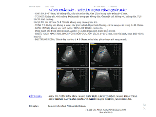
Ultrasound detected hepatosplenomegaly, and thickening of the wall of sigmoid colon with lymph nodes inside abdomen. On the neck, a parotid tumor at the lower pole (tail of parotid) exists on the right side.


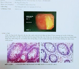
Pathologic result of sigmoid colon was tubular adenoma and slight dysplasia.

Gastroendoscopy revealed an ulcer at lesser curvature and edema of gastric corpus.

Core biopsy of the parotid tumor proved Warthin 's tumor of the right parotid with pathologic result.


ABSTRACT

A male doctor 69 year-old patient with sputum cough for one week.  He got a submandibular mass in suspecting lymphoma or parotid tumor. But on the neck, ultrasound revealed parotid tumor that was concluded a Warthin 's tumor with pathological result of core biopsy.

Morever the doctor patient suffers from a benign tumor of sigmoid colon, gastritis with ulcer and hepatosplenomegaly, and fortunatly gets over the lymphoma haunting.

References

1. Warthin Tumor: Papillary Cystadenoma Lymphomatosum "occurs in the tail of the parotid in aged men". [DeGowin's Diagnostic Examination, 2004].


2. 


Saturday 23 July 2022

CASE 645: ULTRASOUND BUTTERFLY in EMERGENCY ROOM, MEDIC MEDICAL CENTER , HCMC, VIETNAM

ABSTRACT

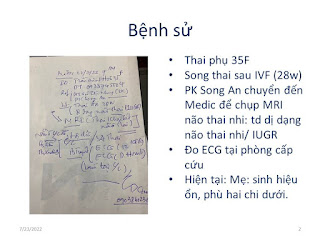
A 35 year-old female patient with twin pregnancy by IVF is in ER of Medic Center that goes through a examimation of check-up by Butterfly ultrasound, and D-heart  for her EKG of 2 fetus and mother heart.
The brains of fetus 26ws and 29ws on fetus ultrasound are normal, but it exists a doubt of club foot for one fetus.
MRI detects fetus brains normal.
Amniocentesis thinks about an heterozygosity of chromosome 18.


Thursday 21 July 2022

CASE 644: HEPATIC ABSCESS due to PERFORATED CHOLECYSTITIS with STONES, Dr DƯƠNG NGỌC THÀNH, Dr LÊ THANH LIÊM, Dr NGUYỄN HỮU QUỐC, Dr TRẦN THỊ HỒNG VÂN, Dr VÕ NG THÀNH NHÂN, Dr PHAN THANH HẢI, MEDIC MEDICAL CENTER, HCMC, VIETNAM

 A 56 year-old female patient suffered from epigastric pain for one month with out fever. WBC 18,880/L , hCRP 129.5mg/L.

Ultrasound of abdomen revealed an abscess of liver that came from a cholecystitis due to stone. Gall bladder wall thickening was perforated that connected to the abscess. And there were stones in CBD and cystic duct.


Abdominal CT confirmed an abscess of liver due to perforated cholecystitis with biliary stones [segment IV- V].  It existed perforation of gall bladder, and stones of commune bile duct and cystic duct.


A medicinal regime was tried for 5 days.


MRI was done before a surgical treatment.


Endoscopic surgery at first but it did to change to open surgery to take away the hepatic abscess, that was in GB bed. Inflammed GB adhered to liver, mesentery and duodenum that has been dissected difficultly. 

Surgeons performed partial cholecystectomy, and Kehr drainage after removing stones in CBD and in cystic duct.


DISCUSSION and CONCLUSION:


Hepatic abscess due to perforated cholecystitis with biliary stone is still a rare entity. Ultrasound could detect successfully cholecystitis due to biliary stone  [84-97 % sensitive, and 95-97% specific] that seems to be higher than CT or MRI does.

Saturday 16 July 2022

CASE 643: MUCUS SECRETION versus ENDOLUMINAL TRACHEA TUMOR, LÊ HỮU LINH MD, MEDIC MEDICAL HCMC, VIETNAM

A 63 year-old male patient goes to the doctor as bloody sputum coughing for some days with feeling of suffocation. There was no pathological sign or symptom of other organs. Inflammed blood tests, and coagulation tests were in normal range. 


Chest CT scan in a local hospital showed a small nodule at the posterior wall of the trachea which was suspected a tracheobronchitis or a papilloma of the trachea. 

His wife and his familial doctor also want having a bronchoscopy for the patient at MEDIC.


3 days later, patient no longer spitted bloody sputum, and completely seems to be healthy. The pulmonologist decided to repeat chest CT and virtual bronchoscopy for the patient. Chest CT at MEDIC showed tracheal lesion disappeared.

DISCUSSION:

Characteristics to detect a mucus secretion in trachea:

       Small size.

       At posterior wall of trachea (supine position during CT scan procedure).

       Small air shadow within the nodule suggesting mucus secretion.

       It will be transformed or disappeared after coughing.


CONCLUSION:

Mucus secretion in trachea can be mistaken with endoluminal trachea tumor.

Their characteristics on chest CT should be put under caution. And a repeated CT scan made after coughing may help detecting a mucus secretion. Thanks of that we could avoid unnecessary invasive procedure for patient. 

Reference case: 

A case with chest CT showed multiple nodular lesions that were both upper lobes, indicated a pulmonary tuberculosis. And it existed a small nodule at posterior wall of trachea. After coughing, repeated CT scan showed the nodule of trachea disappeared.


Friday 15 July 2022

CASE 642: ZENKER' S DIVERTICULUM, Dr PHAN THANH HẢI, Dr NGUYỄN TUẤN CƯỜNG, Dr LÊ HỮU LINH, MEDIC MEDICAL CENTER, HCMC, VIETNAM

 A  67 year-old male patient with multinodular goiter in reexamination.

Ultrasound Medic revealed a cystic lesion with air inside that was in posterior of right thyroid lobe. The lesion may be mimicking a calcified thyroid nodule in right lobe.


CT with CE confirmed the cyst captured contrast media which is in connection to esophagus on the right side. A Zenker's diverticulum was noted.


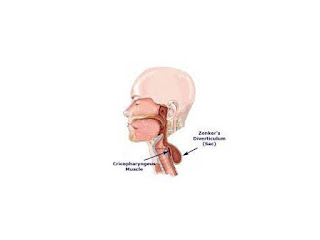
A Zenker's diverticulum is an outpouching that occurs 
at the junction of the lower part of the throat and the
upper portion of the esophagus. The pouch forms 
because the muscle that divides the throat from 
the esophagus, the cricopharyngeal (CP) muscle,
 fails to relax during swallowing. 
https://www.mountsinai.org › zenkers-diverticulum
DISCUSSION:
Zenker's diverticulum is always on left side of the anterior 
neck.
In our case the lesion belongs to right side; and at first 
sight, it looks like a calcified thyroid nodule. But 
we could see some comet tail artifacts below 
the lesion and hence, it does notexist the posterior 
shadowing artifact like in case of a calcified lesion.
CONCLUSION:
We should take different diagnosis for any thyroid lesion 
nearby the esophagus, especially before performing a
 FNAC for a thyroid lesion.


Thursday 7 July 2022

CASE 641: SMALL BREAST TUMOR, Dr JASMINE THANH XUÂN, Dr PHAN THANH HẢI, Dr NẠI THỊ HƯƠNG THOANG, Dr TRẦN THỊ HỒNG VÂN, Dr HỒ CHÍ TRUNG, MEDIC MEDICAL CENTER, HCMC, VIETNAM


A 46 yo female patient goes to Medic center in breast ultrasound screenning.
Breast ultrasound detects an [4x10mm] echo mass, irregular border, inclined axis, with microcalcifications on the right breast.
The right breast mass comes from a tubular breast with microcalcifications inside.


There is not vascular signal in the right breast mass  on Doppler ultrasound.
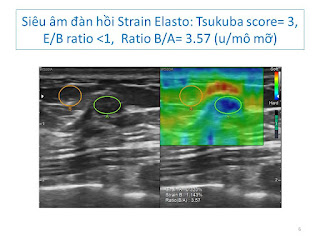
Elastoultrasound strain score 3, ratio B/A=3.57.


Mammography= On right breast it exists a mass # 10 mm, high density, blurre border with microcalcication foci ingathering.


Breast MRI  with gado= Mass of right breast with high signal on T2W2 and low on T1W1, non captured CE, and some breast cysts both 2 sides.
Axillary lymph nodes are inflammed nodes.

Breast thermography: Nothing abnormal detected,  due to it is a small tumor.


Result of core biopsy of the right breast tumor= Invasive breast carcinoma of no special type, grade 2.

 
CONCLUSIONS=

Small size  breast tumor <10mm  may  be revealed early in yearly screenning.

Size, location, characteristic findings will be informed with multimodalities of diagnostic imaging= ultrasound, MRI, thermography and core biopsy.

Pathohistological result is appropriate evident for  breast tumor diagnosing.