Total Pageviews

Monday 20 December 2021

CASE 619 : ABDOMINAL AORTIC DILATATION, Dr PHAN THANH HẢI, Dr TRẦN THỊ THANH NGA, Dr VÕ NGUYỄN THÀNH NHÂN, Dr NGUYỄN THÀNH ĐĂNG, MEDIC MEDICAL CENTER, HCMC, VIETNAM

Old male patient 70 yo, with  AAA suspected came to Medic for reexamination.

Abdominal CT with contrast thought about subrenal non dissection AAA, diameter # 29x32mm, with intramural aortic thrombus and  aortic wall plaques. Left iliac artery in dilatation with plaque and ulcer of vascular wall.


But Doppler color ultrasound showed a dilatation of subrenal abdominal aorta  # 60x18mm with thombus that narrows 30% of  lumen and aliasing artifacts into.


Later MRI of abdomen without Gadolinium confirmed a dilatation of abdominal aorta in 2 sections, the last one with plaques in subrenal part of aorta . Left commun iliac artery with plaques is also in dilatation.


MRI of Medic Center could perform vascular imaging without Gadolinium enhancement.

Friday 12 November 2021

CASE 618: SMALL UTERUS LEADS TO SUSPICION TO CEREBRAL TUMOR, Dr LE DINH VINH PHUC, Dr PHAN THANH HAI, MEDIC MEDICAL CENTER, HCMC, VIETNAM


Female tennager 13 yo from midland region of middle Vietnam go to Medic for a checkup. 

In general ultrasound  a small uterus was detected so inducing a endocrinological problem but therer is no clue about. 


For year got pain of her right leg and foot at gym without abnormal on X-ray films. From 6 months till now 3 right hand fingers, foot and fingers are involuntary in flexion and weakness feelings. Then for later 3 months her chief complaints are plenty drinking, polyuria, headache, space out, more sleeping and drowsiness. But she got no fever nor blurred vision.


MRI brain was performed and a germinoma tumor was detected above hypophysis,hypophysis and basal ganglion on right side.


Sunday 31 October 2021

CASE 617: PROSTATE on SWE TRUS with BIPLAN PROBE, Dr NGUYEN MINH THIEN, Dr PHAN THANH HAI, MEDIC MEDICAL CENTER, HCMC, VIETNAM

Male patient 64yo with fever and voiding discomfort for month.

Digital rectal examination detected a big prostate with a hard nodule on right side.

On MRI there were a 22x15mm hard node of right lobe and another one, 12x16mm, on the left of prostate which concern neoplastic foci , but  PSA=1.75 ng/mL;  F/T=20%


At Medic, an SWE elastography TRUS with biplane probe was done


On TRUS B-mode,  peripheral area of right lobe exists a 16x18 mm hypoechoic nodule, not well-limited,  hypervascular and irregular capsule of right lobe.

On strain elastography, nodule is harder than transitional area while surrounded area of right lobe is softer than the nodule.

On 2D SWE, stiffness of nodule on right side has got a value of 50 kPa [mild hard] while lesion of transitional area of left lobe of 47 kPa.


By via  transperineal a biopsy of prostate was perfomed and histopathological result is TB of prostate and subacute prostatitis.


Conclusion: 

SWE TRUS with biplan probe helps imaging lesions of prostate with more  information to plan for treatment better.


Saturday 30 October 2021

CASE 616: INFECTED SCROTAL SKIN, Dr LE TU PHUC, Dr LE VAN TAI, Dr PHAN THANH HẢI, MEDIC MEDICAL CENTER, HCMC, VIETNAM

A 74-year-old male patient presented to our clinic because of pain and swelling in the right scrotum for 10 days. The scrotum became larger, harder and pus drained out of the scrotum about 3 days before the ultrasound examination.


About 3 months ago, he underwent transurethral resection of the prostate (TURP) and was infected with COVID-19 in the postoperative period. After the COVID isolation, the patient did not show any symptoms of infection.


On ultrasound of the scrotum, we found scrotal edema, thickening, interstitial fluid and gas between the scrotal skin layers. Gas spreads anteriorly to the right pubic tubercle and posteriorly to the base of the penis near the anus.


Gas was not seen in the left scrotum, in the spermatic cord, in the skin of the abdomen and on the buttocks and thighs. No fistula from the rectum was found.


Due to the patient's recent urinary tract surgery, urinary catheterization, history of diabetes, gas and fluid in the right scrotal skin. We therefore assumed diagnosis of Fournier's gangrene.


The paitent was transferred to surgery department after and treated with debridement surgery in combination with antibacterial and detoxification therapy. He improve well till now.


Getting well at hospital discharge.


Saturday 17 July 2021

CASE 614: ELASTOGRAPHY ULTRASOUND for a Case of CKD, Dr PHAN THANH HẢI, Dr NGUYỄN NGHIỆP VĂN, MEDIC MEDICAL CENTER, HCMC, VIETNAM

Male patient 38 yo, follow-up for CKD for years and now preparing hemodialysis by via kidney machine.
Ultrasound B-mode shows hyperechoic pattern of parenchymal and pyelonephritic structure both 2 kidneys but dimensions and vascular Doppler remain in normal ranges.


Elastography ultrasound for kidney was performed on ARFI SIEMENS S 2000, VTQ= 1.4-2.22m/sec


and SWE Supersonics machine= 22-24kPa.

As the results, values of renal parenchymal stiffness increase slightly in 2 machines with 2 different technics of elastography ultrasound.

DISCUSSIONS:
1/ Shear wave elastography and ARFI technic may be a low-cost way to provide additional diagnostic information in CKD.
2/ Our case with increased  values of both  ARFI for CKD from 1.4--2.22m/s and SWE measurements = 22-24kPa that proves a heterogeneicity of kidney, and the  complex parenchymal stiffness which may lead to fibrosing of kidney  in progress.

REFERENCES:
Acoustic Radiation Force  Impulse Imaging for Evaluation of Renal Parenchyma Elasticity in Diabetic Nephropathy, Cemil Goya et al. AJR 2015; 204:324–329
Applications of acoustic radiation force impulse quantification in chronic kidney disease: a review, Liang Wang, Ultrasonography 2016;35:302-308
Shear wave elastography in chronic kidney disease: a pilot experience in native kidneys , Samir et al. BMC Nephrology (2015) 16:119


Sunday 2 May 2021

CASE 613 : BILATERAL BREAST CANCER, Dr PHAN THANH HAI, Dr NGUYEN HUU QUOC, Dr TRAN LAM

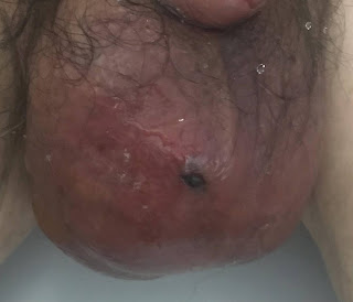
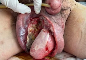
Female patient 82 yo, herself detected a right breast mass that had been in traditional management, but it was getting slowly bigger. Now the right breast is swollen and hemorrhage.


Ultrasound detected tumors at right and left breast. Noted BI-RADS 5 with metastase lymph nodes of right breast tumor, and BI-RADS 4A for the left one.


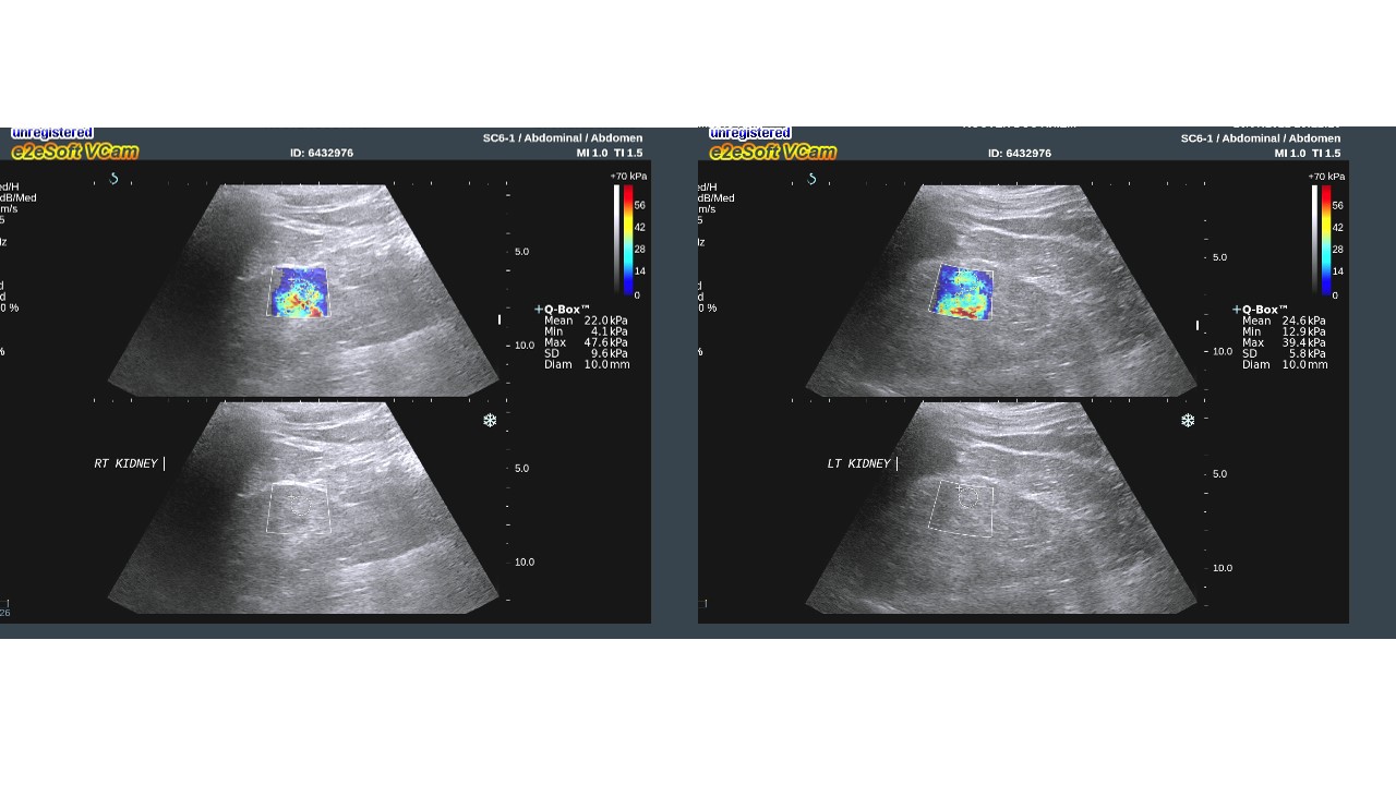
Ultrasound findings:
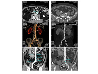
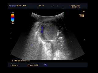
Right breast = Lesion with mass effect is at 1/2 lateral breast, heterogenous, multiloculated, solid inside with multicalcified foci and neoplastic vessels. Limits of right pectoralis muscle and the lesion is not clear. This lesion invades subcutaneous fatty tissue and right breast skin . Because of big size tumor it can not examined posteriorly the breast tumor. 

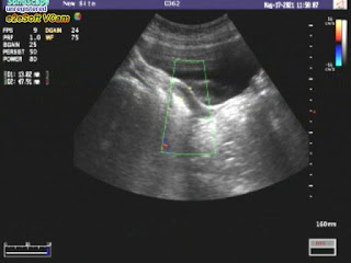
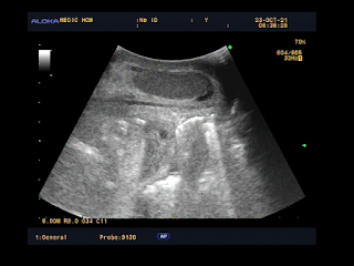
Left breast= Many cystic structures with calcified foci, the biggest one is at 3 o'clock and far from center about 3 cm.
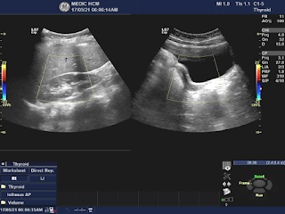
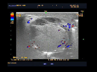
Many lymph nodes loss hilus with microcalcification exist in axillary areas both 2 sides and in right supraclavicular fossa.
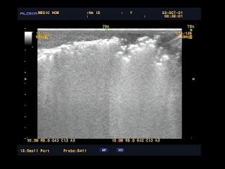
Comet tail artifacts at the lung base both 2 sides.


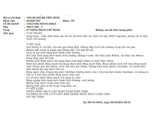
MSCT with CE  also detected right  breast tumor and lymph nodes.


CT= Right breast lesion is heterogenous tissue condensation, multiloculated invades breast skin and deformes right breast, highly captured CE, # 61.3 x 80.2mm. Some right axillary lymph nodes # 11mm. There is pulmonary fibrosis  and pleural thickening at the right lung base.
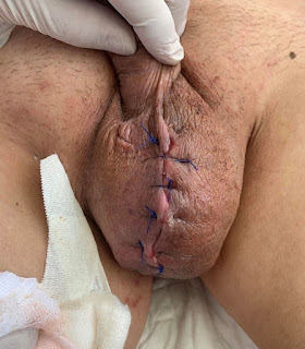
Wait for surgery.