Total Pageviews

Wednesday 1 July 2020

CASE 589: TB OF TESTIS, Dr PHAN THANH HẢI-Dr LÊ THANH LIÊM, MEDIC MEDICAL CENTER, HCMC, VIETNAM


Male 28yo, with swelling and scrotal pain in thrombophlebitis management and spermatic vein thrombosis for 2 months but nothing change that a hospital in HCM city made decision to surgery because of not ruling out a sarcoma?

US and MRI cannot rule out a spermatic tumor.


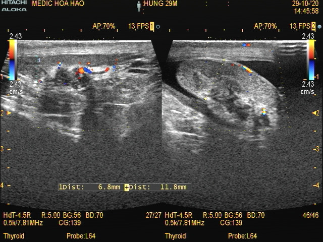
At Medic Center utrasound detected edema of epidydimal head, hypervascular #36x24mm with some calcium nodules, scrotal skin edema and small amount of  fluid in scrotum while seldom revealed lymph nodes that are poor echoic  in necrosis and calcified  at left neck=10-31mm that made  thought about TB abscess of left epidydimis


Blood tests: WBC 10.900 / mL;  CRP 13.63 mg/L; AFP 1.94 ng/ml; BetaHCG blood < 0.2  mUI/ml.


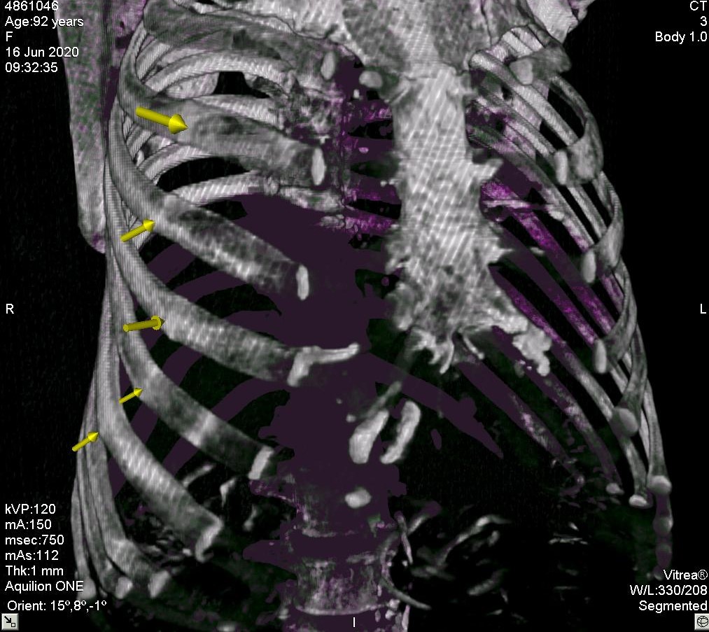
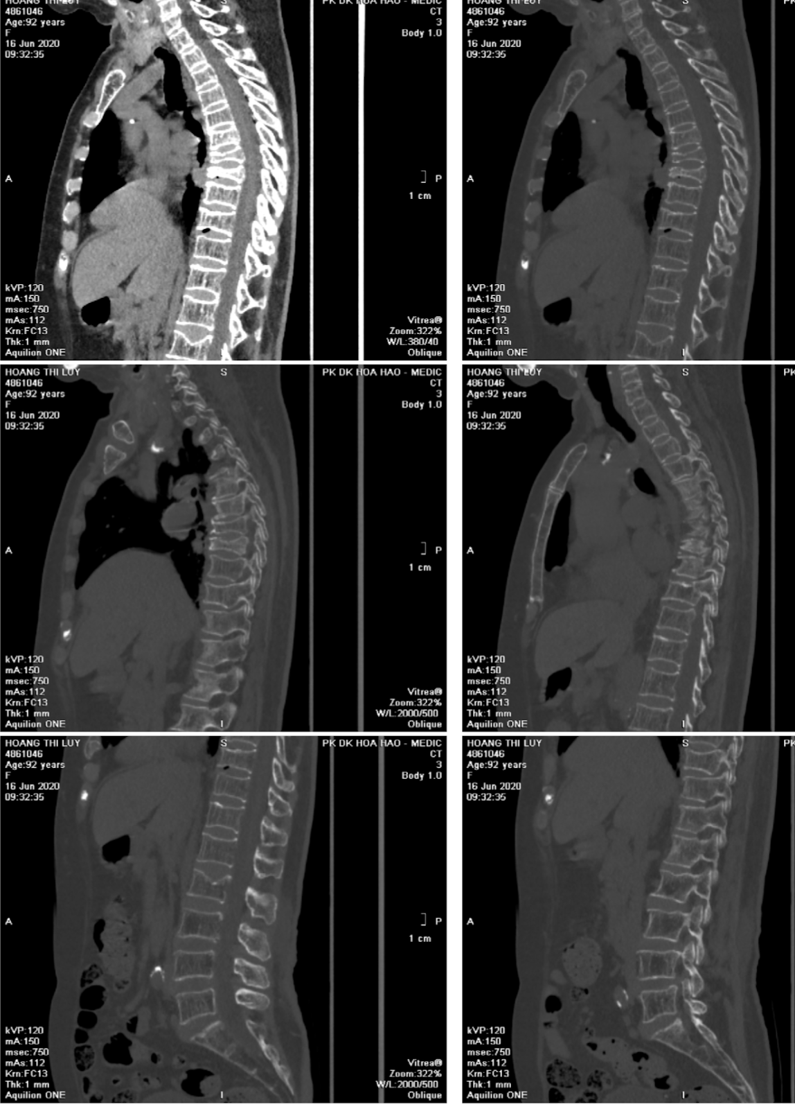
Chest X-Rays detected fibrotic lesion in right subclavian area and suspected TB lesion of right lung.


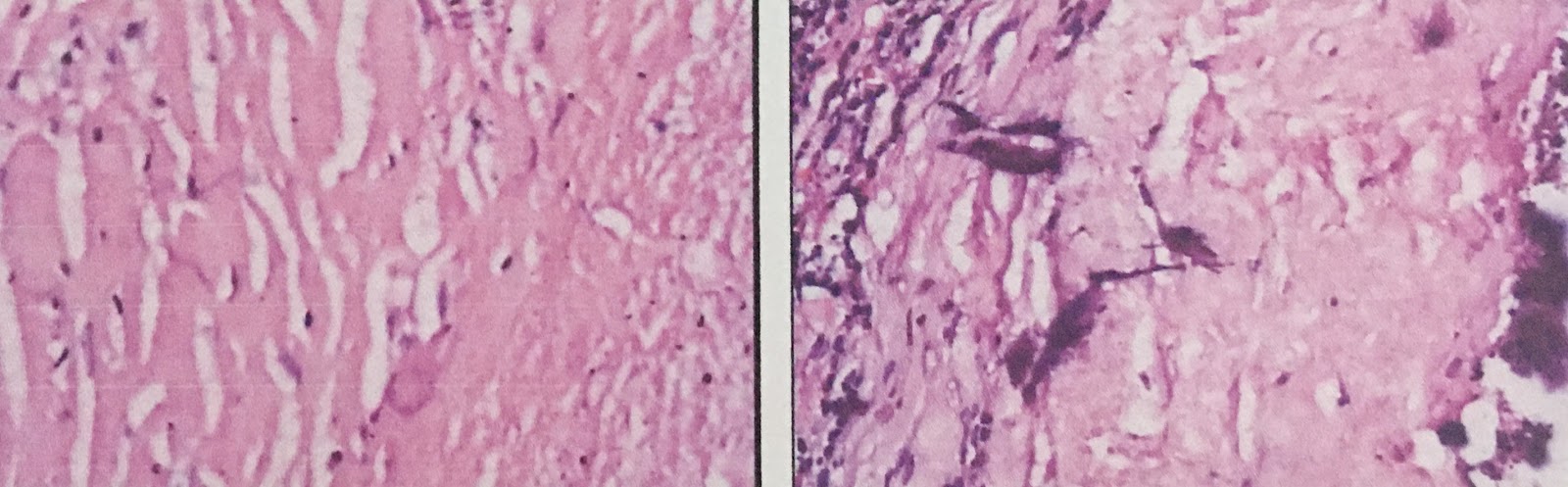
FNAC for left neck lymph node thinks about TB node.


Pulmonary and TB PNT hospital suspected TB testis and peripheric nodular disorders.


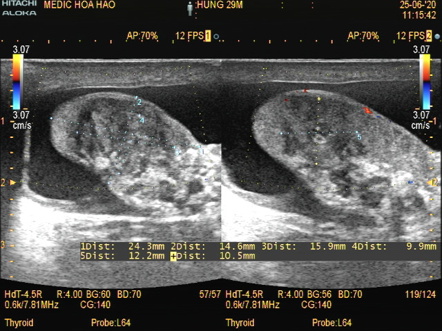
For 4 months of TB treatment, on ultrasound in Medic Center, head of epidydimis decreases volume #24x16mm, hyperechoic pattern, non hypervascular irrigation with existing a small abscess of 16x11mm, and scrotal skin slightly thickend with small amount of fluid in scrotum.


Decreasing of volume of left neck lymph nodes =10-29mm.

Post TB therapy course 9 months
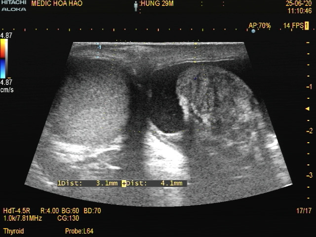
Small scare of epidydimis, normal testicular vein. Neck nodes reduce the sizes.


Tuesday 30 June 2020

CASE 588:THIGH MUSCLE THICKENING [TMT] AFTER FEMUR BONE FRACTURE, Dr PHANTHANH HẢI, Dr LÊ THỊ THANH THẢO, Dr PHAN THANH HẢI PHƯỢNG, MEDIC MEDICAL CENTER, HCMC, VIETNAM


Female 86yo. Right femur neck fracture due to falling for 2 months.

X-Rays = Right neck femur bone fracture, Garden 4. Severe osteoporosis on Osteogram and calcaneus ultrasound.


MSK ULTRASOUND=

Old fracture of right femur neck with existing callus, non continuous rough surface and edema soft tissue around without hematoma.


Ultrasound for sarcopenia by mesurement of Thigh Muscle Thickening (TMT)=
 R= 12mm/ L=23mm.
Noted decreasing of  right thigh muscle [rectus and mediale femoris] volume.


Sunday 7 June 2020

CASE 587 : POCUS for A CASE of CHEST PAIN, Dr PHAN THANH HẢI , Dr LÊ THANH LIÊM, MEDIC MEDICAL CENTER, HCMC, VIETNAM


Female 92 yo, complaint of a right chest pain and subcostal pain for 5 days that gets more painful when coughing and moving. In emergency examination, she is in consciousness and well contact.
Pouls and blood tension are normal


EKG PoC at bed :Ischemic myocardial  regions in lateral anterior, lateral high  and diaphragmatic of heart.


Chest X-Rays PoC (in supine) results: Cardiopathy due to atherosclerosis, nothing abnormal detected of lungs, pleural and thoracic cage, elevated right diaphragm.


Lung Ultrasound PoCUS 
No pneumothorax proved by existing sliding sign (+).


No pleural effusion


Abdomen Ultrasound PoCUS = No free fluid, no findings of contusion of solid organs = liver, spleen, pancreas, kidneys.


Thoracic wal ultrasound PoCUS= Light fracture of 3rd rib anterior arcade without deplacement, and soft tissue around slight edema.


CONCLUSION:

 PoCUS at home helps ruling out dangerous conditions like pneumothorax, hemopleuresis, hemoperitoneum, solid organ contusions. PoCUS may evaluate painful points and decide on site (at home) appropriate managements. In face- to - face  contact, PoCUS  may help patient coming down and getting out of anxiety in emergency. 


UPDATE


Friday 29 May 2020

CASE 586: PLACENTA ACCRETA or not, Dr PHAN THANH HẢI, Dr JASMINE THANH XUÂN, Dr VÕ HIẾU THÀNH, Dr THÙY MAI, MEDIC MEDICAL CENTER, HCMC,VIETNAM.

Female 25 yo, PARA  0020,  with 01 surgery for extrauterine pregnancy,  now suspected placenta accreta in 37,5 weeks of pregnancy by ultrasound in Hung vuong hospital.


Ultrasound and MRI in Medic= placental accreta in small part in left angle of uterine fundus.

Ultrasound showed a thin part of uterus with a poor placenta part nearby that made thinking about placenta accreta.


MRI= It is difficult to see muscular layer of left side of uterine fundus that may be invaded abnormally by placenta accreta.


Finally, results of cesarean surgery shows a normal placenta.


Discussion= Wrong thinkings of ultrasound due to abnormal of uterus post op: at the site of the late surgery,  the poor echogeneicity of  part of placenta made thinking about placenta accreta. However, it exists non Doppler signal at this site, so nothing proved for an evident of placenta accreta. 

Nota= 

There were some ultrasound findings of placeta accreta=
 - vascular lacunae  in small and large size in placenta with hypervascular and   turbulent flows
 - loss of hypoechoic line in posterior of placenta
 - abnormal of wall of bladder due to invasion by placenta 

Friday 22 May 2020

CASE 585: MRI VENOGRAPHY of GALIEN VEIN ANEURYSM, Dr PHAN THANH HẢI, Dr NGUYỄN THÀNH ĐĂNG, MEDIC MEDICAL CENTER, HCMC, VIETNAM.

Female child  4yo, having a vascular cystic lesion had been revealed  by T C D ultrasound into her brain without symptom.


Per transcranial sonography (TCD) it exists   a cystic lesion #6x8 mm with venous spectral pattern and aliasing that appeares nearby the cerebral troncus. The cystic lesion seems to be no changing of its size for 4 years.


MRI venography of brain performed to confirm the results of TCD ultrasound,  and detect clearly an aneursym of  Galien vein in upper space  of  tentorium cerebelli  in the female child brain.


MRI venography has a important role to confirm an aneurysm of Galien vein for the female child.