Total Pageviews

Friday 29 May 2020

CASE 586: PLACENTA ACCRETA or not, Dr PHAN THANH HẢI, Dr JASMINE THANH XUÂN, Dr VÕ HIẾU THÀNH, Dr THÙY MAI, MEDIC MEDICAL CENTER, HCMC,VIETNAM.

Female 25 yo, PARA  0020,  with 01 surgery for extrauterine pregnancy,  now suspected placenta accreta in 37,5 weeks of pregnancy by ultrasound in Hung vuong hospital.


Ultrasound and MRI in Medic= placental accreta in small part in left angle of uterine fundus.

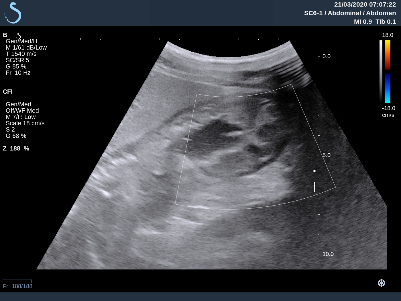
Ultrasound showed a thin part of uterus with a poor placenta part nearby that made thinking about placenta accreta.


MRI= It is difficult to see muscular layer of left side of uterine fundus that may be invaded abnormally by placenta accreta.


Finally, results of cesarean surgery shows a normal placenta.


Discussion= Wrong thinkings of ultrasound due to abnormal of uterus post op: at the site of the late surgery,  the poor echogeneicity of  part of placenta made thinking about placenta accreta. However, it exists non Doppler signal at this site, so nothing proved for an evident of placenta accreta. 

Nota= 

There were some ultrasound findings of placeta accreta=
 - vascular lacunae  in small and large size in placenta with hypervascular and   turbulent flows
 - loss of hypoechoic line in posterior of placenta
 - abnormal of wall of bladder due to invasion by placenta 

Friday 22 May 2020

CASE 585: MRI VENOGRAPHY of GALIEN VEIN ANEURYSM, Dr PHAN THANH HẢI, Dr NGUYỄN THÀNH ĐĂNG, MEDIC MEDICAL CENTER, HCMC, VIETNAM.

Female child  4yo, having a vascular cystic lesion had been revealed  by T C D ultrasound into her brain without symptom.


Per transcranial sonography (TCD) it exists   a cystic lesion #6x8 mm with venous spectral pattern and aliasing that appeares nearby the cerebral troncus. The cystic lesion seems to be no changing of its size for 4 years.


MRI venography of brain performed to confirm the results of TCD ultrasound,  and detect clearly an aneursym of  Galien vein in upper space  of  tentorium cerebelli  in the female child brain.


MRI venography has a important role to confirm an aneurysm of Galien vein for the female child. 

Thursday 14 May 2020

CASE 584: INFANTILE MESENCHYMAL HAMARTOMA IN LIVER, Dr PHAN THANH HẢI, Dr LÊ THANH LIÊM, Prof NGUYỄN SÀO TRUNG, MEDIC MEDICAL CENTER, HCMC, VIETNAM.


Male child 3 months old, well feeding and weight .
Dr Truong Dinh Khai [Children Hospital N2] detected liver lesions with high level of AFP, suspected hepatoblastoma. But Wako tests= AFP 6,388.4; AFP L3 and DCP in normal values.

At Medic, ultrasound thought about infectious lesions in right lobe #56x53mm, solid, septation with cystic appearance. 


MRI (Gado)= Right lobe of liver lesions may belong to mesenchymal sarcoma, AFP  got down <2,000.


Operation for removing liver tumor.
Macro and microscopic specimens with results are Mesenchymal Hamartoma in liver.
The specimen composes of cords of normal hepatocytes with loose cellular parenchyme, congestive blood vessels, hyalynized fibrous tissue.

2 months post op selective hepatectomy, in reexamination check-up, regular weight gain normaly # 8kg of 5.5 months old, AFP downed at 44 ng/mL (last time 74ng/mL). Not detected relapse tumor. 

Conclusion :

Hepatic Mesenchymal Hamartoma is a rare benign tumor in children. Tumor appeares in big cyst, septated  or solid matter with small cysts.  Hepatic mesenchymal sarcoma is a different diagnostic item with asthenia, invasion to vessels, biliary obstruction.


Saturday 18 April 2020

CASE 583: ANTRUM Linitis Plastica, Dr PHAN THANH HẢI, Dr LẬP, Prof LÊ QUANG NGHĨA, MEDIC MEDICAL CENTER, HCMC, VIETNAM


Man 38yo, nurse, could not eat, lost 13 kg for 3 months.

With 3 times of gastroendoscopy and biopsy  he was treated as gastritis with Hp-positive  (see gastro endoscopy and biopsy result) at Medic HCMC  


His report of abdominal ultrasound examination pointed out antral thickening like a black ring (see US1 crossed- section; US 2 longitudinal scanning).


MSCT CE report: gastritis with antral thickeking (see CT-scan). 


At Medic blood tests CEA, CA 72-4, CA 19-9 are all normal. 

Abdominal x-rays with barium showed typical Linitis Plastica of the antrum. 


This patient was admitted to Binh Dan hospital emergency department to undergo endoscopic US which revealed antral thickening more than 2 cm.   


Total gastrectomy and replacement by small intestine were performed (see macro: TOTAL GASTRECTOMY)


Microscopic report post op is Adenocarcinoma that invaded to intramuscular wall and going  metastasis to lymph nodes.


Monday 24 February 2020

CASE 582: INSULINOMA, Dr PHAN THANH HAI, Dr HUYNH TRAC LUAN, MEDIC MEDICAL CENTER, HCMC, VIETNAM.

WOMAN  25YO  with  HYPOGLYCEMIA COMA MANY TIMES  IN EMERGENCY  HOSPITAL.   

AT MEDIC, BLOODTESTS:  
FASTING GLUCOSE  (FPG) = 2.4  (N=3.9-5.9 mmol/mL)    
Insuline=   71.3  ( N= 2.6-24.9 microUI/mL) 
C-PEPTIC FASTING = 7.45 ng/mL (N =1.1-4.4 mUI/mL)
BLOOD TEST RESULTS SUSPECTED INSULINOMA.  
ULTRASOUND of  PANCREAS IS NORMAL  BUT SMALL ACCESSORY  SPLEEN EXISTS (US 1, US 2, US 3).


MSCT SPECTRAL CONTRAST of PANCREAS  NON DETECTED PANCREAS TUMOR BUT CLEARLY VIEWING ACCESSORY SPLEEN (CT1/CT2).


MRI of PANCREAS   DETECTED  SMALL TUMOR AT THE TAIL of PANCREAS #1.5 CM  SUSPECTED  INSULINOMA.


Operation removed small tumor at the tail of pancreas  (ope, macro1, macro2).


MICROSCOPIC REPORT IS  ENDOCRINE PANCREATIC  ADENOMA  (INSULINOMA).


CONCLUSION = CASE OF TYPICAL INSULINOMA IN CLINICAL   BUT ULTRASOUND and CT CANNOT DETECT THIS SMALL TUMOR, EXCEPT MRI.

Reference    CT perfusion of pancreas  detected insulinoma.


Wednesday 19 February 2020

CASE 581: RETROPERITONEAL GIANT LIPOMA, Dr PHAN THANH HAI, Dr TRAN THI NGA, MEDIC MEDICAL C, HCMC, VIETNAM.

Man 35 yo with distention of abdomen slowly  after liposuction intra abdominal  wall.


Ultrasound of abdomen detected big mass from epigastric to hypogastric area,  hypovascular and echostructure like fatty tissue. 

US 1: the mass near the liver border  hypovascular
US 2: crossed-section at epigastric  this mass  around  AMA.  
US 3: crossed-section at umbilic area,  aorta and IVC no replaced.
US 4: sagittal middle abdomen   aorta and vci  
ultrasound  report is big lipoma intra abdomen.


MSCT CE    
CT1  CROSSED SECTION  L KID  DEPLACEMENT  
CT2  FRONTAL VIEW   BIG LIPOMA  WITH HU  IS 30UI
CT3  FRONTAL SECTION THIS TUMOR IS RETROPERITONEUM DEPLACEMENT BOWEL TO RIGHT SITE
CT4  3D VIEW  HS L KIDNEY ROTATION  


CT REPORT IS RETROPERITONEUM LIPOMA  
OPERATION  REMOVED 7 KG  TUMOR  AND MICROSCOPIC REPORT IS LIPOMA.


REFERENCE CASE