Total Pageviews

Saturday 18 April 2020

CASE 583: ANTRUM Linitis Plastica, Dr PHAN THANH HẢI, Dr LẬP, Prof LÊ QUANG NGHĨA, MEDIC MEDICAL CENTER, HCMC, VIETNAM


Man 38yo, nurse, could not eat, lost 13 kg for 3 months.

With 3 times of gastroendoscopy and biopsy  he was treated as gastritis with Hp-positive  (see gastro endoscopy and biopsy result) at Medic HCMC  


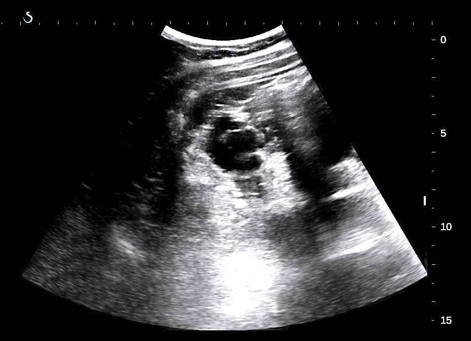
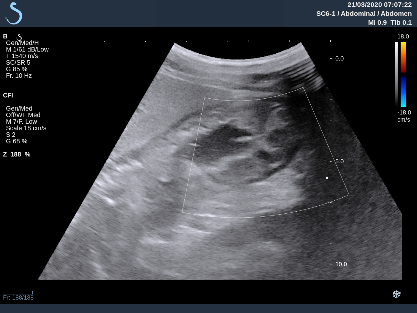
His report of abdominal ultrasound examination pointed out antral thickening like a black ring (see US1 crossed- section; US 2 longitudinal scanning).


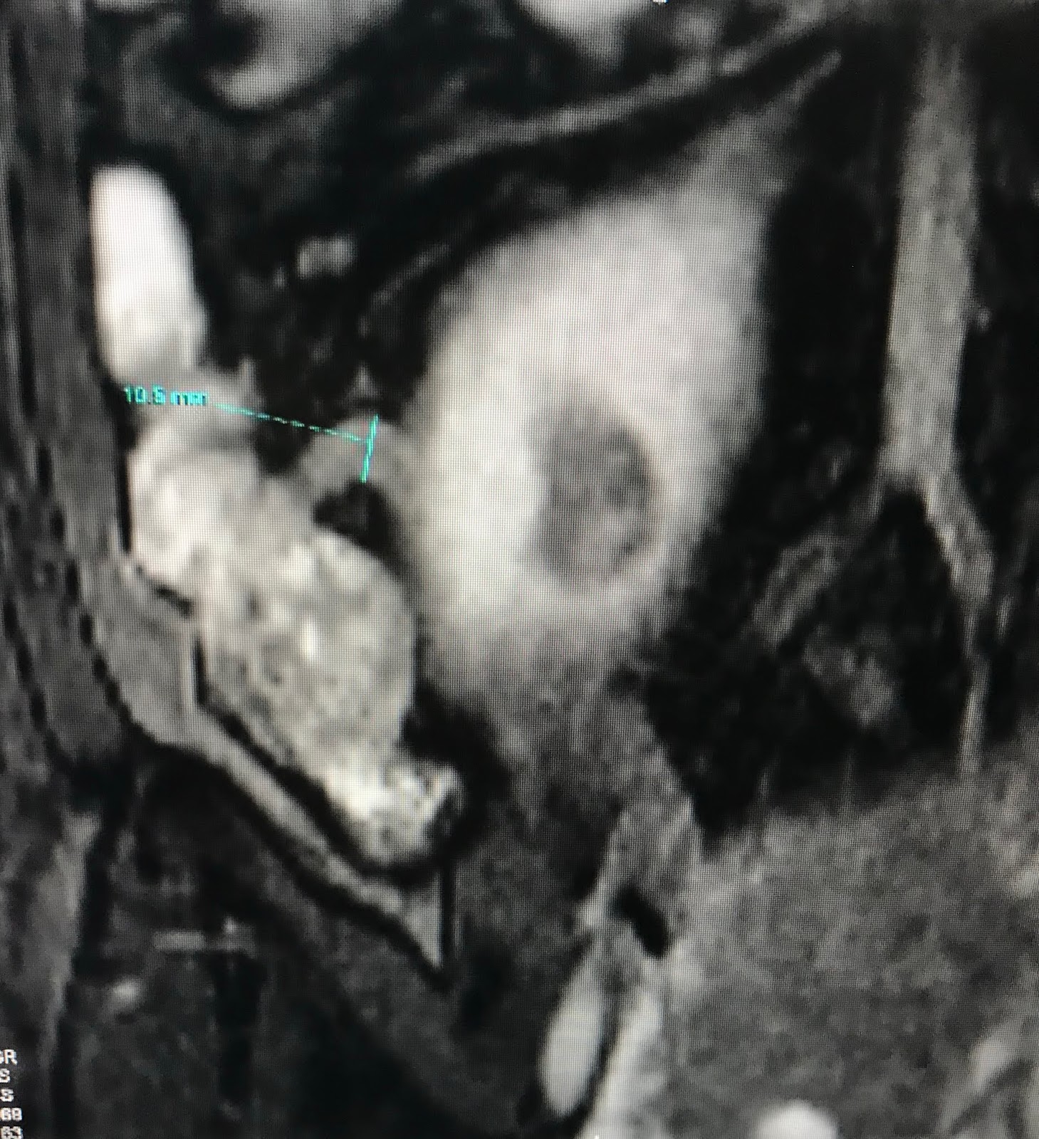
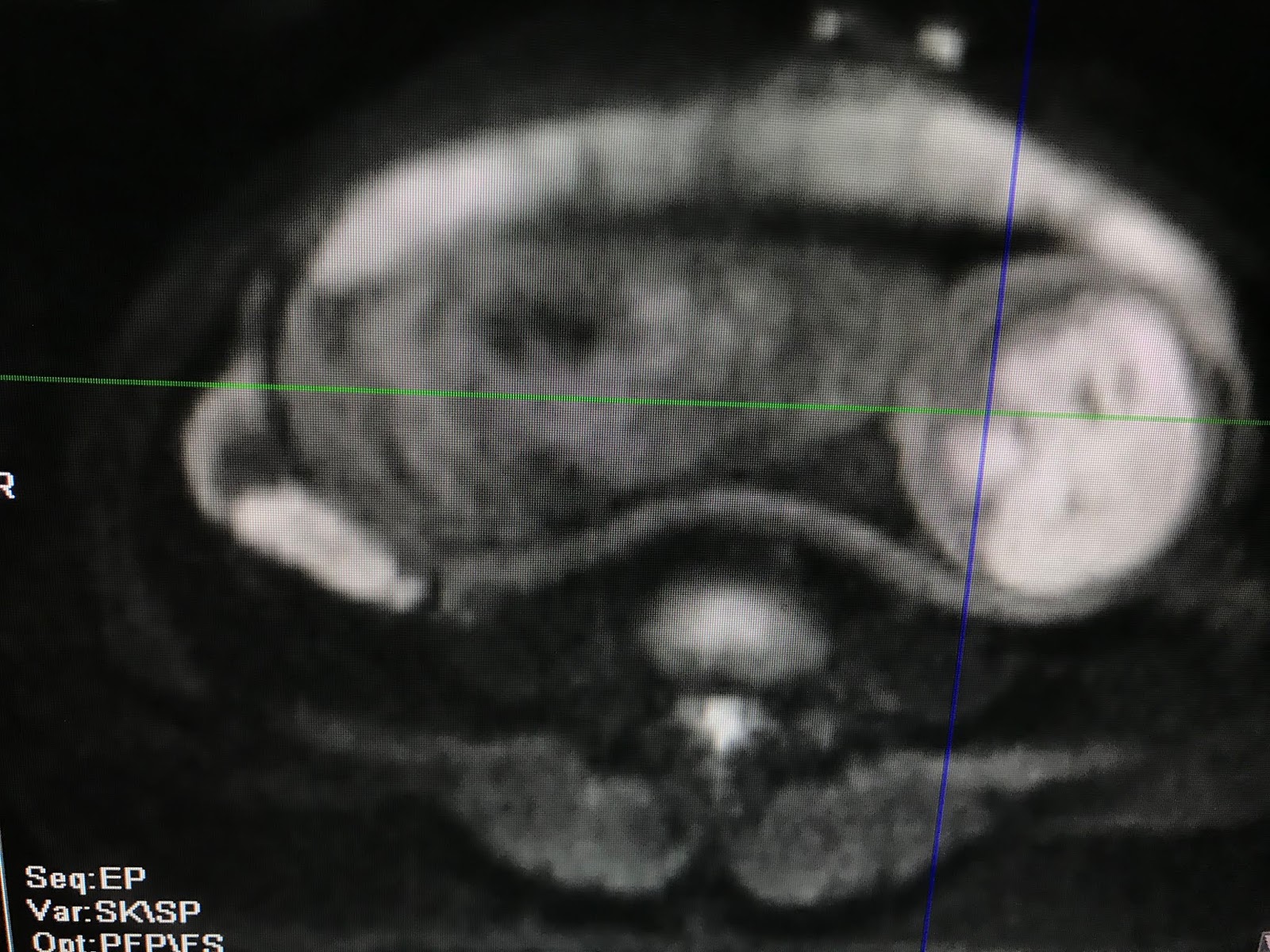
MSCT CE report: gastritis with antral thickeking (see CT-scan). 


At Medic blood tests CEA, CA 72-4, CA 19-9 are all normal. 

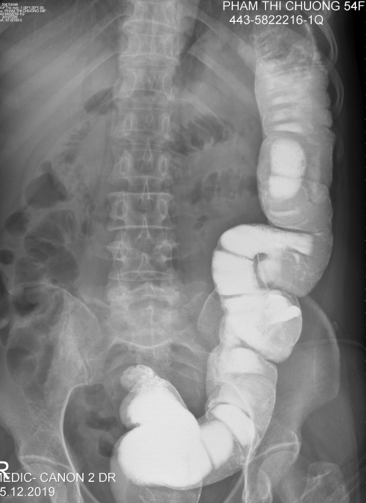
Abdominal x-rays with barium showed typical Linitis Plastica of the antrum. 


This patient was admitted to Binh Dan hospital emergency department to undergo endoscopic US which revealed antral thickening more than 2 cm.   


Total gastrectomy and replacement by small intestine were performed (see macro: TOTAL GASTRECTOMY)


Microscopic report post op is Adenocarcinoma that invaded to intramuscular wall and going  metastasis to lymph nodes.


Monday 24 February 2020

CASE 582: INSULINOMA, Dr PHAN THANH HAI, Dr HUYNH TRAC LUAN, MEDIC MEDICAL CENTER, HCMC, VIETNAM.

WOMAN  25YO  with  HYPOGLYCEMIA COMA MANY TIMES  IN EMERGENCY  HOSPITAL.   

AT MEDIC, BLOODTESTS:  
FASTING GLUCOSE  (FPG) = 2.4  (N=3.9-5.9 mmol/mL)    
Insuline=   71.3  ( N= 2.6-24.9 microUI/mL) 
C-PEPTIC FASTING = 7.45 ng/mL (N =1.1-4.4 mUI/mL)
BLOOD TEST RESULTS SUSPECTED INSULINOMA.  
ULTRASOUND of  PANCREAS IS NORMAL  BUT SMALL ACCESSORY  SPLEEN EXISTS (US 1, US 2, US 3).


MSCT SPECTRAL CONTRAST of PANCREAS  NON DETECTED PANCREAS TUMOR BUT CLEARLY VIEWING ACCESSORY SPLEEN (CT1/CT2).


MRI of PANCREAS   DETECTED  SMALL TUMOR AT THE TAIL of PANCREAS #1.5 CM  SUSPECTED  INSULINOMA.


Operation removed small tumor at the tail of pancreas  (ope, macro1, macro2).


MICROSCOPIC REPORT IS  ENDOCRINE PANCREATIC  ADENOMA  (INSULINOMA).


CONCLUSION = CASE OF TYPICAL INSULINOMA IN CLINICAL   BUT ULTRASOUND and CT CANNOT DETECT THIS SMALL TUMOR, EXCEPT MRI.

Reference    CT perfusion of pancreas  detected insulinoma.


Wednesday 19 February 2020

CASE 581: RETROPERITONEAL GIANT LIPOMA, Dr PHAN THANH HAI, Dr TRAN THI NGA, MEDIC MEDICAL C, HCMC, VIETNAM.

Man 35 yo with distention of abdomen slowly  after liposuction intra abdominal  wall.


Ultrasound of abdomen detected big mass from epigastric to hypogastric area,  hypovascular and echostructure like fatty tissue. 

US 1: the mass near the liver border  hypovascular
US 2: crossed-section at epigastric  this mass  around  AMA.  
US 3: crossed-section at umbilic area,  aorta and IVC no replaced.
US 4: sagittal middle abdomen   aorta and vci  
ultrasound  report is big lipoma intra abdomen.


MSCT CE    
CT1  CROSSED SECTION  L KID  DEPLACEMENT  
CT2  FRONTAL VIEW   BIG LIPOMA  WITH HU  IS 30UI
CT3  FRONTAL SECTION THIS TUMOR IS RETROPERITONEUM DEPLACEMENT BOWEL TO RIGHT SITE
CT4  3D VIEW  HS L KIDNEY ROTATION  


CT REPORT IS RETROPERITONEUM LIPOMA  
OPERATION  REMOVED 7 KG  TUMOR  AND MICROSCOPIC REPORT IS LIPOMA.


REFERENCE CASE


Sunday 16 February 2020

CASE 580: COLON INTUSSUSCEPTION, Dr PHAN THANH HẢI, Dr LÊ ÐÌNH TÍN, MEDIC MEDICAL CENTER, HCMC, VIET NAM.

WOMAN 54yo DURING TREATED HYPERTHYROIDISM  SHE HAD GOT DIARRHEA WITH MUCUS MANY TIMES AND PAIN IN LLAQ.


EMERGENCY ULTRASOUND OF ABDOMEN  DETECTED  ONE MASS INTRA SIGMA COLON  WITH TARGET SIGN IMAGE ( US 1, US 2), SIZE 5 CM  INDUCED  COLON INTUSSUSCEPTION    AND THYROID HYPERVASCULAR PATTERN  ( US 3).


FOR DIAGNOSTIC PERFORMED COLON ENEMA WITH BARYUM (X-RAY 1, X-RAY 2)  SHOW ONE MASS INTRA SIGMA COLON  5CM IN SIZE.


MSCT OF ABDOMEN:   THIS MASS IS HYPODENSITY HU  LIKE LIPOMA .


ENDOSCOPY OF COLON  DETECTED TUMOR BUT BIOPSY RESULT IS  MUCOSAL INFLAMATION.


WAIT FOR TREATED HYPERTHYROIDISM STABLE TO SURGERY IN SAFETY.

Operation  detected  tumor of descending colon near  splenic angle. Macroscopic is like lipoma, wait for microscopic report.


MICROSCOPIC REPORT IS  LIPOMA    


REFERENCE   CASE REPORT  


CONCLUSION  =  LIPOMA INTRACOLON  INDUCED COLON INTUSSUSCEPTION.

Friday 24 January 2020

CASE 579: ACUTE APPENDICITIS in PREGNANCY, Dr PHAN THANH HAI, MEDIC MEDICAL CENTER, HCMC, VIETNAM.

Woman 33 yo in pregnancy. PARA 3003 # 24 weeks WITH pain in RLAQ 24 HOURS AGO
  
OBSTETRIC ULTRASOUND PROVED NORMAL PREGNANCY IN  24WKS WITHOUT UTERUS CONTRACTION IN TOCOMETRY (PHOTO TOCO).


EMERGENCY ULTRASOUND  DETECTED APPENDIX IN BIG  LUMEN DUE  TO DILATATION (US 1, US 2, US 3).


BLOOD TESTS:  WBC RISED 9.8K  NEUTRO 40%, CRP 56..
MRI  WITHOUT GADO  DETECTED   BIG  LUMEN DILATED APPENDIX (MRI 1, MRI 2, MRI 3).


ENDOSCOPIC  OPERATION REMOVED ACUTE APPENDICITIS.

  
SUMMARY = ACUTE APPENDICITIS IN PREGNANCY IS DIFFICULT TO DIAGNOSE;   US AND MRI  CONTRIBUTING  TO DIAGNOSE  IN DIFFICULT CASE.

REFERENCE : CASE 548, APPENDICITIS IN PREGNANCY  (MEDIC ULTRASOUND CASE 548  PUB 01 MAY-2019)