Total Pageviews

Sunday 14 October 2018

CASE 518: FINGER TUMORS, Dr PHAN THANH HẢI, Dr NGUYỄN NGHIỆP VĂN, Dr TRẦN THỊ THANH NGA, Dr LÊ THÔNG LƯU, MEDIC MEDICAL CENTER, HCMC, VIETNAM.

Woman 20yo with right hand  having many tumors at fingers,  slow growth, difficult movement of flexion ( see foto   (right hand  dorsal view:   tumor at first finger, finger number 3  and  number 5)  color skin  is changed,   soft in palpation and  no pain.


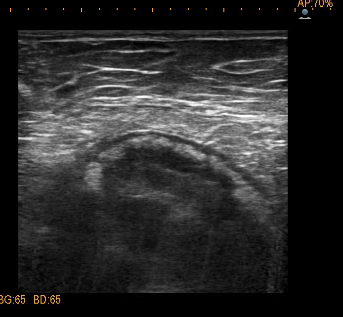
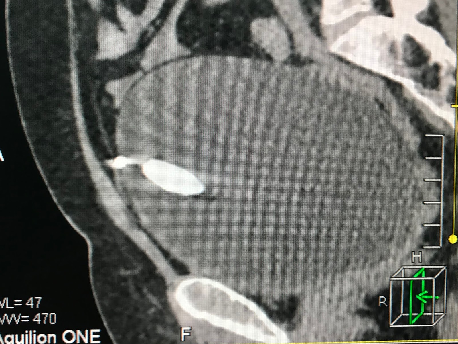
Ultrasound of  this tumor by linear probe 11 MHz : US 1:  tumor solid  hypoechoic, ellypsoid ;
 2 -3cm, central necrosis; crossed section ( US 2 ) with more vascular supplying and not fixed to bone.


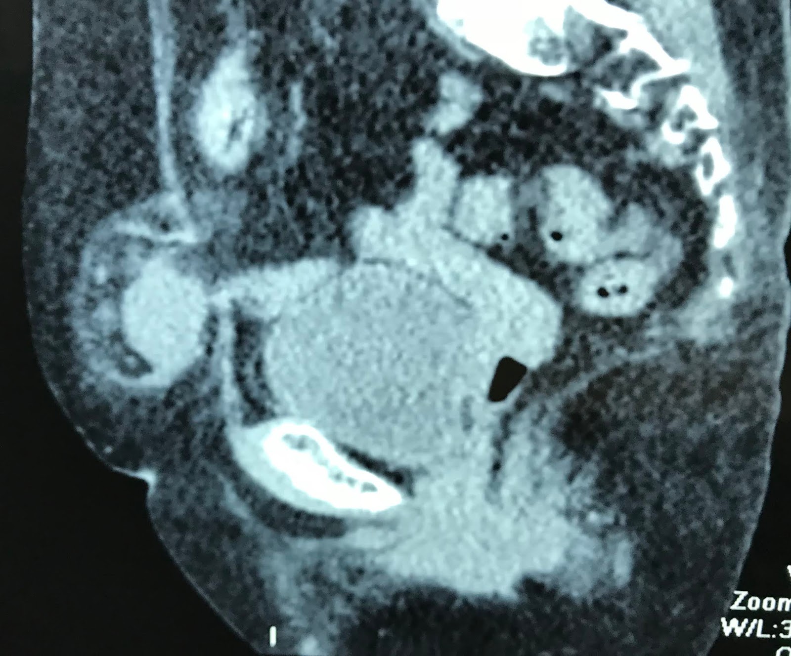
Another tumor only in right hand not related to joint .
Clinical history: she was being treated in hospital as  hemangioma,   but  sonologist said  it is geant cell tumor of the tendon sheath.

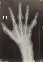
X-Rays films of the right hand: No erosion of the bone.
Abnormal atrophic metatarsal number 4. Typical cavernous hemangioma and phleboliths.


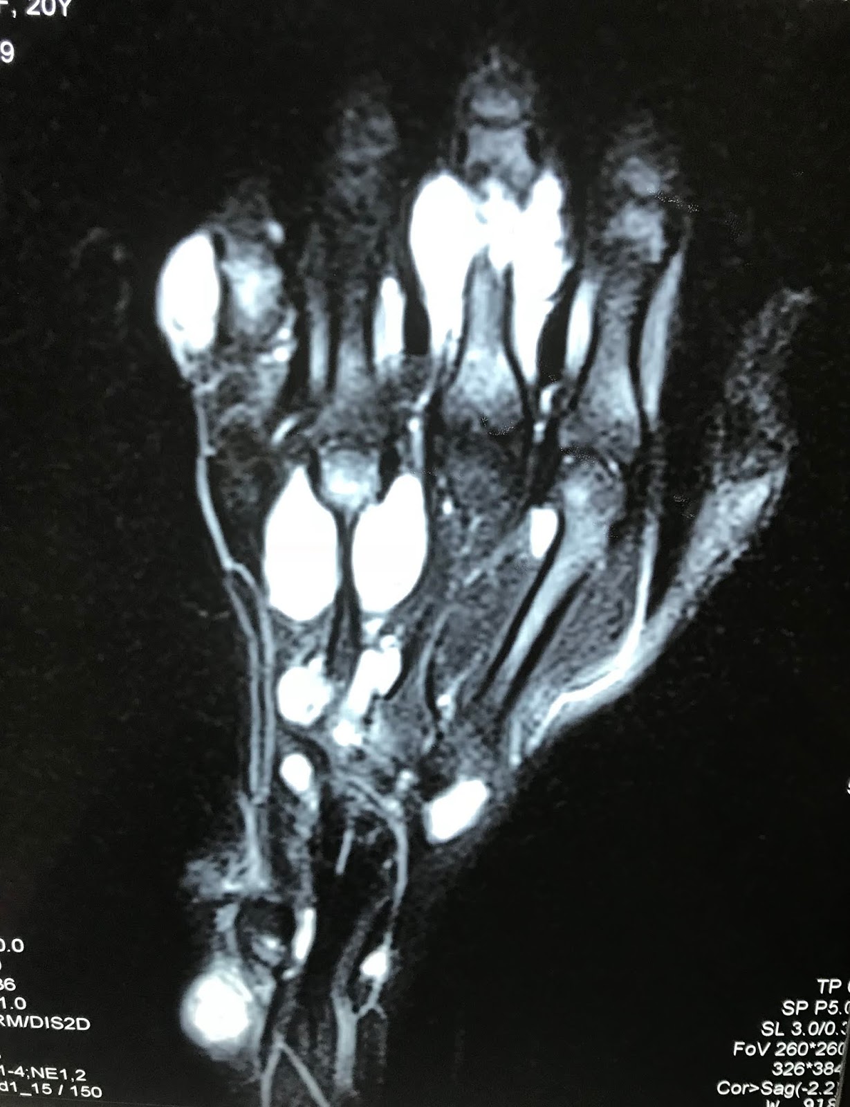
MRI OF RIGHT HAND   SHOWS MULTIPLE TUMORS.


Operation removed one small tumor at first finger.


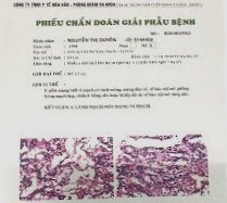
Histology report is benign capillary hemangioma, perycyte hemangioma of fingers.


Monday 8 October 2018

CASE 517: HCC WITH LOW AFP, Dr PHAN THANH HẢI, MEDIC MEDICAL CENTER, HCMC, VIETNAM.


MAN 52 YO with HBV INFECTED and   EPIGATRIC PAIN.   
ULTRASOUND CHECK UP DETECTED  LEFT LIVER TUMOR, SIZE 3.5 CM   HYPOECHOIC (US1,US2  )   BLOOD TETS: HBsAg POSITIVE, AFP 15ng/mL.


CT SCAN LIVER =  CT 1  NON CE , CT2, CT3   TRIPHASES CE , CT4 FRONTAL VIEW , CT5  SAGITTAL VIEW.

RADIOLOGIST REPORT IS  HCC.


MRI PRIMOVIST = MRI 1 DWI ,  MRI 2  T1, MRI 3 GADO INPUT,   MRI 4 GADO OUT,   MRI 5  FRONTAL VIEW.


BLOOD TEST AGAIN=  WAKO TEST  AFP-L3  RISING 16.4 %.


SUMMARY=  MAN 52 YO HBV INFECTED
WITH AFP NEGATIVE   BUT ULTRASOUND DETECTED TUMOR AND CT CE , MRI GADO   STUDYING THIS TUMOR.   WAKO TEST IS  POSITIVE  WITH L3 RISING. HOW ACCURATE DIAGNOSTIC OF   ALL FACILITIES?
OPERATION IN THE PLAN of LEFT HEPATECTOMY.

Robotic operation for  left hepatectomy.   See  macro  tumor with white area.

Microscopic with histo immunostaining  is  undiffentiated HCC.


Friday 5 October 2018

CASE 516: PANCREAS TUMOR, Dr PHAN THANH HAI, Dr TRUONG DINH KHAI, MEDIC MEDICAL CENTER, HCMC, VIETNAM


Girl  10yo with  epigastric pain.
Ultrasound of abdomen detected  one mass of 8cm  at the body of pancreas, cystic  structure,  well bordered  ( US 1,  US 2, US 3),  no lymph node around.


MSCT with CE=   this mass is from the tail of pancreas,  inhomogeneous structure (CT1 CT 2, CT 3).


Blood tests  are normal.
Operation for resection of this tumor  [see macro].

CT  SHOWS THIS TUMOR A SOLID TUMOR BUT ULTRASOUND SCAN FINDING IS CYSTIC.

Summary:  Girl 10 yo  with big mas at the pancreas,  structure is mixed solid and cystic,  the most common  is  solid pseudocystic papillary tumor.
Histology result is pancreas pseudopapillary neoplasia.


REFERENCE:PDF.

Friday 28 September 2018

CASE 515: VENTRAL HERNIA, Dr PHAN THANH HẢI, Dr LÊ THỊ THANH THẢO, MEDIC MEDICAL CENTER, HCMC, VIETNAM.


Woman  78 yo,  cesarian operation for  50 years, with, 2 days ago,  pain at left umbilical area  and everytime  coughing, a swollen mass appearing at the painful site.  

Ultrasound  of  this mass: 
US 1=  longitudinal scan at pelvis  near umbilicus, detected one subcutaneous mass. 


US 2 = left lateral scanning of middle line, shows  the tearing site of rectus muscle and a hernia goes out from it.


US 3 =  linear scanning of  this mass that was builded by great omentum and fluid.


US 4 = thickening of the bowel wall.


Emergency abdomen CT scanning:
CT  1:  crossed section  of  this mass  


CT 2 :  sagittal scanning of this mass


CT 3:  frontal section.


Emergency operation by  ventral hernia with past history of  cesarian operation 
detected  omentum and one bowel loop in ischemia but not necrosis yet.


Repaired this hernia by a mesh prosthesis. And timing is about in 4 hours from diagnostic to operation.

Tuesday 25 September 2018

CASE 514: IUD PENETRATING TO URINARY BLADDER, Dr PHAN THANH HAI, Dr JASMINE THANH XUAN, Dr NGUYEN MINH THIEN, Dr NGUYEN TUAN VINH, MEDIC MEDICAL CENTER, HCMC, VIETNAM.


Woman 49yo with pain after urinary miction. PAST HISTORY of BEING PUT T - SHAPE IUD 20 YEARS BEFORE.
ULTRASOUND of PELVIS  DETECTED BIG URINARY BLADDER STONE ( US1)

XRAY of PELVIS  SHOWs THIS STONE  WITH IUD  INSIDE   ( X-RAYs film).

ULTRASOUND  WITH CDI  FINDs OUT TWINKLING  ARTIFACTS  WITH  COMET TAIL SIGN in GREEN AND RED COLORS ( US 2, US 3).


MSCT of  PELVIS  :
 CT 1=  THE  METALIC IUD  INTRA UB WALL.

CT 2:  SAGITTAL VIEW .

CT 3: FRONTAL VIEW : THE  IUD  INTRA UB WALL.

ENDOSCOPY DETECTED THE STONE IN VAULT OF UB.

Operation removed a big stone intra urinary bladder.


CONCLUSION =  IUD  PENETRATING TO UB WALL AND FORMATION OF STONE.

IUD Migration in MEDIC Hoa Hao