Total Pageviews

Saturday 4 August 2018

CASE 505: LEG ISCHEMIA, Dr PHAN THANH HẢI, MEDIC MEDICAL CENTER, HCMC, VIETNAM.


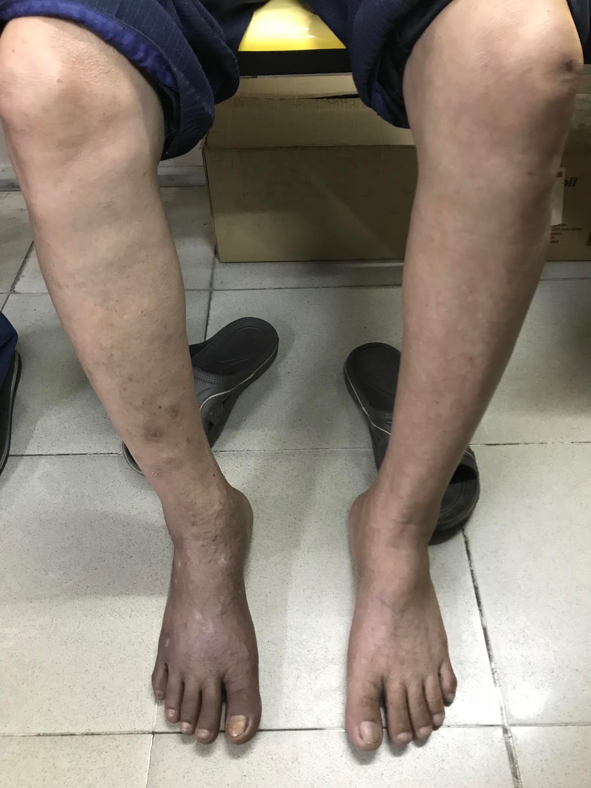
Man 70 yo in claudication with  pain at right leg and right foot changes in dark skin (photo).


Vascular ultrasound:   
US 1=  normal left femoral artery and vein.


 US 2=  right femoral artery and vein in stenosis.


 US 3=  in middle of right  thigh cannot find out superficial artery.


 US 4= high flow of right dorsalis pedis artery.


Thermography  shows the right leg in hypothermia.


MSCT  angio of the leg arteries:  

CT 1=  big cysts in right/left kidneys and  abdominal aorta with sclerosis plaque.


CT 2=  right  superficial femoral artery is in obstruction. 


CT 3=  small anastomosis at the level of right thigh.


Diagnosis =  obstruction of right superficial femoral artery  with many sites of deep and superficial anastomosis of femoral artery.


DSA  shows  complete obstruction of right superficial femoral artery (DSA 1)


DSA 2 = small anastomosis.


Operation removed blood clot and revascularization of the right leg. Patient remains well recovery.


Sunday 22 July 2018

CASE 504: PERIHEPATIC ABSCESS DUE TO FISHBONE, Dr PHAN THANH HẢI, Dr LÊ THỊ THANH THẢO, MEDIC MEDICAL CENTER, HCMC, VIETNAM


Woman 43 yo with  epigastric pain  has been treated as gastritis.
Ultrasound detected near  falciform ligamentun of liver  one abscess  with  foreign body long 3 cm  look like  fishbone  ( US1, US 2, US 3  SWE shows  it is very hard, US video).


CT scan no CE shows this fishbone is in a perihepatic abscess (CT1). 


One month later  the second CT examination with CE:  CT 2  shows  this abscess is  pulling abdominal wall out like a  hourglass [CT 2,  CT 3  sagital view].


Emergency operation of this abscess retrieved pus with a fishbone.


Saturday 7 July 2018

CASE 503: SUBUNGUAL GLOMUS TUMOR, Dr LÝ HỮU ĐỨC, Dr PHAN THANH HẢI, MEDIC MEDICAL CENTER, HCMC, VIETNAM.


WOMAN  46 YO  WITH  HISTORY  OF  5 YEARS AGO  PAIN AT  FIRST FINGER OF LEFT HAND, SWELLING AND BLEEDDING.
ULTRASOUND  OF THIS FINGER   WITH CDI    US 1   LONGITUDINAL SCANNING   DETECTED  ONE  MASS HYPOECHOIC BUT HYPERVASCULAR.


US 2: CROSSED-SECTION OF THIS MASS UNDER UNGUAL  
 HYPERVASCULAR  OF DISTAL FINGER.


MRI 2  =THIS MASS IS UNDER  UNGUAL   NO  INVASION OF THE BONE.


MRI 3 = CROSSED- SECTION OF THIS TUMOR IS  SUB UNGUAL WITHOUT EROSION OF THE BONE.


OPERATION  REMOVED THIS TUMOR.


 MICROSCOPIC RESULT IS  GLOMUS TUMOR.

Monday 2 July 2018

CASE 502: DIFFUSE SKIN XANTHOMA, Dr DƯƠNG NGỌC THÀNH, Dr LÝ HỮU ĐỨC, Dr PHAN THANH HẢI, MEDIC MEDICAL CENTER, HCMC, VIETNAM.


MAN 32 YO 2 YEARS AGO   BEGINNING AT THE HAND   AND FOOT  RISING SMALL NODULES AND HARD, SIZE 1-2 CM, COULORED  REED TO YELLOW   AND FULL BODY  APPEARED BUT NOT IN THE FACE. SEE PHOTOS ( PHOTO 1  THE BACK ,  PHOTO 2  THE FOOT,  PHOTO 3   THE HAND,  PHOTO 4  IN FOCUS NODULES,  PHPOTO 5  THE SKIN OVER ACHILLUS TENDON.


ULTRASOUND:   US 1=  THE NODULE 1,5 CM  UNDER SKIN   AND FATTY LAYER,   US 2 =  CDI  NO BLOOD TO THIS NODULE,  US 3  ACHILLE TENDON IS  NORMAL .


BLOOD TESTS MADE DIAGNOSING ARE DIABETES AND HYPERCHOLESTEROLEMIA AND RISING TRIGLYCERIDS.

BIOPSY  ( SEE MACRO).  


WHILE WAITING FOR  PATHOLOGY REPORT,
ONE DERMATOLOGIST SAYS IT IS ERUPTIVE  XANTHOMATOSIS.


MICROSCOPIC RESULT IS XANTHOMA.

For one month treatment, xanthomatous nodules reduce # 70%.


Saturday 30 June 2018

CASE 501: BLADDER HERNIA to SCROTUM, Dr LÊ TỰ PHÚC, Dr PHAN THANH HẢI, MEDIC MEDICAL CENTER, HCMC, VIETNAM.

A 51-year-old man with pubic pain for 2 weeks with left scrotum swelling and
 increasing in size for 6 months. He has to  compress the scrotum by hand in order to empty the bladder everytime normal ending of micturition= two-stage micturition: the patient after a first spontaneous voiding, presses the mass and voids again. No other urinary symptoms. There was no history of nausea or vomiting.

Ultrasound scanning detected one sac containing of fluid on the left side of scrotum. This sac with thick wall continued upwards left pubic tubercle and getting smaller like a bird beak. This sac appears bigger when the patient did holding his breath (Valsalva maneuver) and disappered after being compression by hand to voiding.


Retrograde cystography CT revealed a left scrotal hernia with fluid density lesion continuous with left lateral bladder wall.


Diagnosis of left scrotal hernia with bladder as content was made.
Operation for repairing urinary bladder was done. A part of bladder adheres at deep orifice of inguinal canal and overcomes to scrotum by upper branch of pubis.


Have you' d ever seen a case like that before and what do you think?